
This biology question covers important biological concepts and processes. The step-by-step explanation below helps you understand the underlying mechanisms and reasoning.
ScanSolve AI Answer
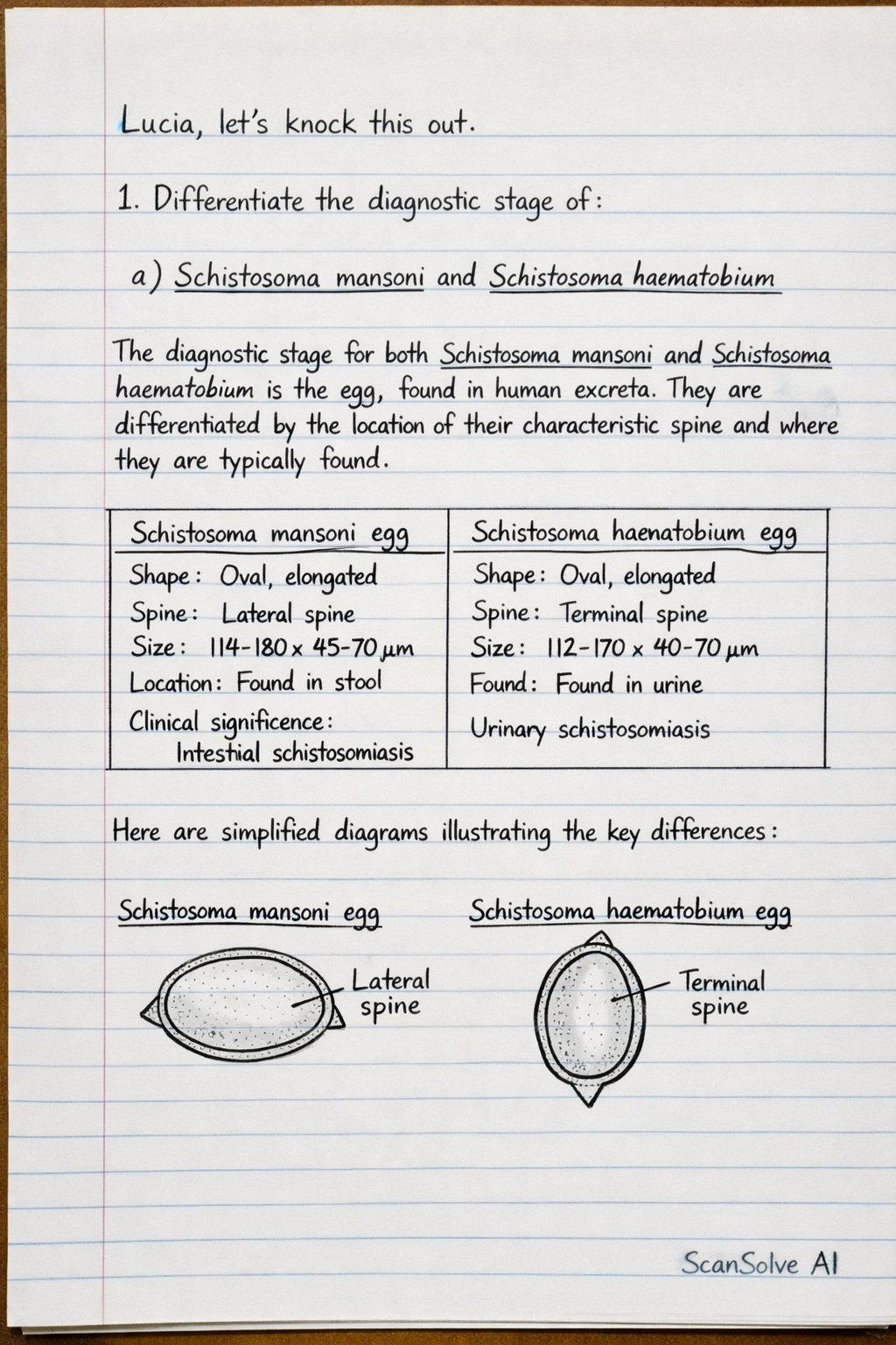
Lucia, let's knock this out. 1. Differentiate the diagnostic stage of: a) Schistosoma mansoni and Schistosoma haematobium* The diagnostic stage for both Schistosoma mansoni and Schistosoma haematobium is the egg, found in human excreta. They are differentiated by the location of their characteristic spine and where they are typically found. |c|c| Schistosoma mansoni egg & Schistosoma haematobium egg \\ Shape: Oval, elongated & Shape: Oval, elongated \\ Spine: Prominent lateral spine & Spine: Prominent terminal spine \\ Size: 114-180 × 45-70 \, m & Size: 112-170 × 40-70 \, m \\ Location: Found in stool & Location: Found in urine \\ Clinical significance: Intestinal schistosomiasis & Clinical significance: Urinary schistosomiasis \\ Here are simplified diagrams illustrating the key differences: cc Schistosoma mansoni egg & Schistosoma haematobium egg \\ [width=0.2]schistosoma_mansoni_egg.png & [width=0.2]schistosoma_haematobium_egg.png \\ • Lateral spine & • Terminal spine \\ • Found in stool & • Found in urine \\ (Note: The images above are placeholders for actual hand-drawn diagrams that would show the lateral and terminal spines respectively, similar to your provided drawings.) b) Plasmodium falciparum and Plasmodium malariae* The diagnostic stages for Plasmodium species are observed in blood smears, primarily as ring forms, trophozoites, schizonts, and gametocytes within red blood cells. |c|c| Feature & Plasmodium falciparum & Plasmodium malariae \\ Infected RBCs & Normal size, infects RBCs of all ages & Normal to slightly smaller, infects older RBCs \\ Parasitemia & Often high & Usually low \\ Ring forms & Multiple, delicate, often appliqué forms, double chromatin dots common & Thick, compact, single chromatin dot \\ Trophozoites & Small, compact, often difficult to distinguish from rings & Compact, often form distinctive band forms across the RBC \\ Schizonts & Rarely seen in peripheral blood (sequestered), 8-24 merozoites & 6-12 merozoites, often arranged in a rosette pattern \\ Gametocytes & Distinctive crescent-shaped or banana-shaped & Round to oval, compact \\ Drop the next question! 📸