
We read your photo as:
Draw and label the diagram of a pseudostratified epithelium
ScanSolve AI Answer
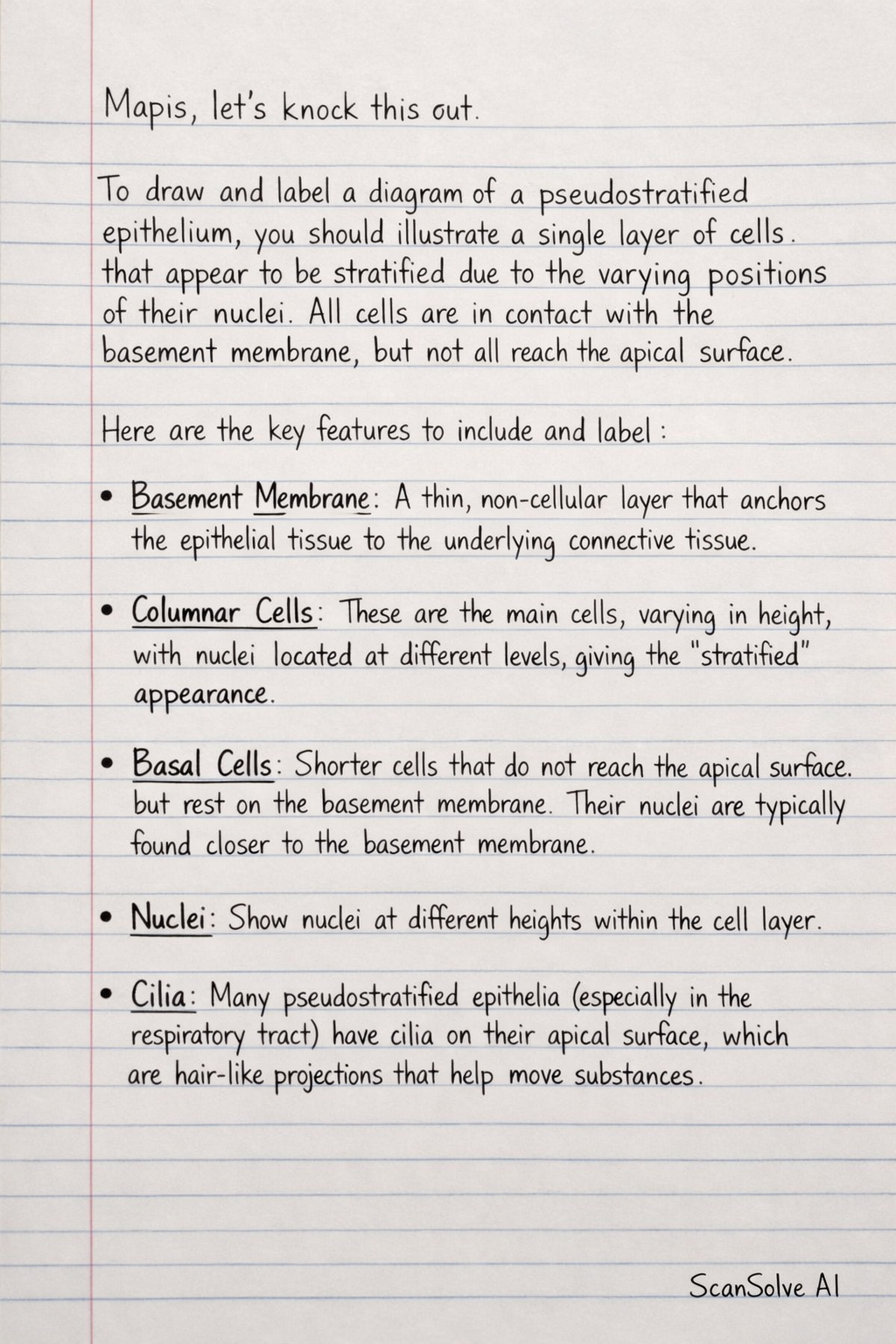
To draw and label a diagram of a pseudostratified epithelium, you should illustrate a single layer of cells that appear to be stratified due to the varying positions of their nuclei. All cells are in contact with the basement membrane, but not all reach the apical surface.
Here are the key features to include and label:
- Basement Membrane: A thin, non-cellular layer that anchors the epithelial tissue to the underlying connective tissue.
- Columnar Cells: These are the main cells, varying in height, with nuclei located at different levels, giving the "stratified" appearance.
- Basal Cells: Shorter cells that do not reach the apical surface but rest on the basement membrane. Their nuclei are typically found closer to the basement membrane.
- Nuclei: Show nuclei at different heights within the cell layer.
- Cilia: Many pseudostratified epithelia (especially in the respiratory tract) have cilia on their apical surface, which are hair-like projections that help move substances.
- Goblet Cells: These are specialized cells often found in pseudostratified ciliated columnar epithelium. They produce and secrete mucus.
A typical diagram would show a row of cells of varying heights, all touching the basement membrane. The nuclei would be scattered at different levels, not in a neat row. Cilia and goblet cells would be present on the apical surface if it's a ciliated type.
Still stuck on this one?
Ask a follow-up, and the answer starts from this question.
More Biology Questions
Handwritten Step-by-Step Solution
Get this as a handwritten image on WhatsApp — like a tutor wrote it for you.
Try on WhatsApp