
We read your photo as:
Identify and label the transverse sections of dicot and monocot stems, distinguishing them based on vascular bundle arrangement.
Biology
ScanSolve AI Answer
Here is the identification and labeling for the transverse sections of the dicot and monocot stems shown in the image.
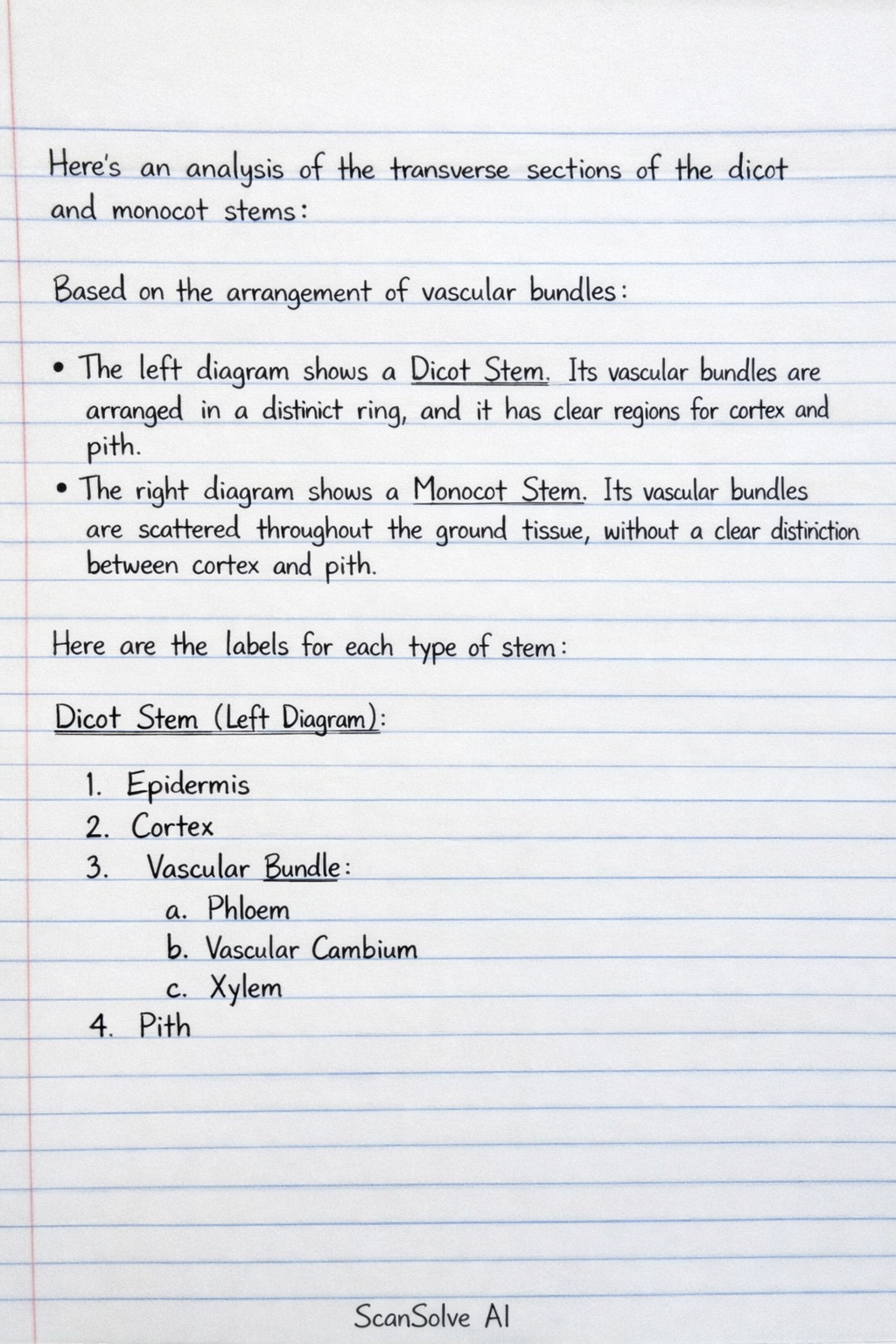
Based on the arrangement of vascular bundles:
- The left diagram shows a Dicot Stem. Its vascular bundles are arranged in a distinct ring, and it has clear regions for cortex and pith.
- The right diagram shows a Monocot Stem. Its vascular bundles are scattered throughout the ground tissue, without a clear distinction between cortex and pith.
Here are the labels for each type of stem:
Dicot Stem (Left Diagram)
- Epidermis: The outermost protective layer, typically a single layer of cells.
- Cortex: Located just beneath the epidermis, extending to the vascular bundles. It consists of:
- Collenchyma: Often found directly under the epidermis, providing flexible support.
- Parenchyma: Ground tissue cells for storage.
- Sclerenchyma: May be present as a cap over the vascular bundles or in other supportive roles.
- Vascular Bundles: Arranged in a ring. Each bundle contains:
- Phloem: Located towards the outside of the stem, responsible for transporting sugars.
- Xylem: Located towards the inside of the stem, responsible for transporting water and minerals.
- Vascular Cambium: (Not explicitly visible at this magnification but present between xylem and phloem) Responsible for secondary growth.
- Pith: The central region of the stem, composed of parenchyma cells, primarily for storage.
- Medullary Rays: Parenchymatous cells located between the vascular bundles, connecting the cortex and pith.
Monocot Stem (Right Diagram)
- Epidermis: The outermost protective layer.
- Ground Tissue: The entire internal region of the stem, composed of parenchyma cells, where the vascular bundles are embedded. There is no distinct cortex or pith.
- Vascular Bundles: Scattered throughout the ground tissue. Each bundle is typically oval-shaped and surrounded by a sclerenchymatous bundle sheath for support. Each bundle contains:
- Phloem: Located towards the periphery of the bundle.
- Xylem: Typically V or Y-shaped, with larger metaxylem vessels and smaller protoxylem vessels, often with a protoxylem lacuna (air cavity).
Was this helpful?
Still stuck on this one?
Ask a follow-up, and the answer starts from this question.
Got a different question?Ask your own question
More Biology Questions
QA suitable caption for the diagram is: Transverse section of a dicotyledonous stem.QSimilarities between monocots and dicots stemQPrepare and examine a transverse section of the stem and root of a dicotyledonous plant under a…QDraw the internal structure of a monocotyledonous root and lable it parts. 2) Draw the internal…QCompare and contrast the root systems and leaf venation of monocots and dicots.
Handwritten Step-by-Step Solution
Get this as a handwritten image on WhatsApp — like a tutor wrote it for you.
Try on WhatsApp