
We read your photo as:
List the subdivisions of the nervous system.
Biology
ScanSolve AI Answer
Step-by-step solution
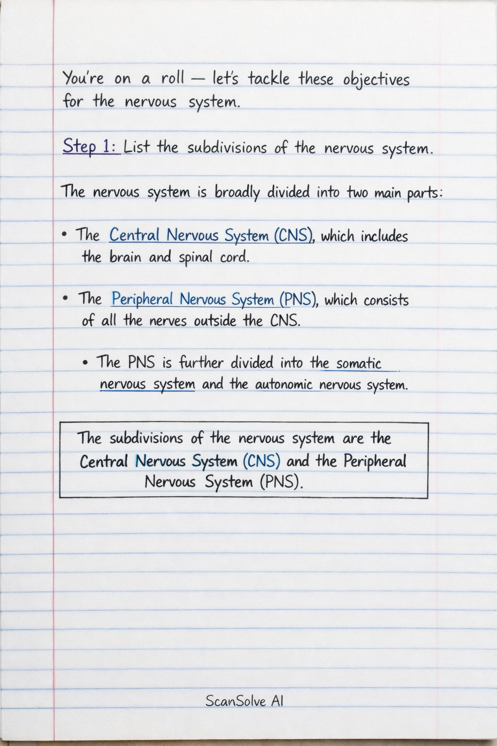
8 stepslet's tackle these objectives for the nervous system.
List the subdivisions of the nervous system.
The nervous system is broadly divided into two main parts:
- The Central Nervous System (CNS), which includes the brain and spinal cord.
- The Peripheral Nervous System (PNS), which consists of all the nerves outside the CNS. The PNS is further divided into the somatic nervous system and the autonomic nervous system.
Define grey matter, white matter, nucleus, ganglion, tract, and nerve.
- Grey matter consists primarily of neuron cell bodies, dendrites, unmyelinated axons, and neuroglia. It is involved in processing information.
- White matter is composed mainly of myelinated axons, which transmit signals rapidly. The myelin sheath gives it its white appearance.
- A nucleus (in the CNS) is a cluster of neuron cell bodies within the central nervous system.
- A ganglion (in the PNS) is a cluster of neuron cell bodies in the peripheral nervous system.
- A tract (in the CNS) is a bundle of axons within the central nervous system that share a common origin, destination, and function.
- A nerve (in the PNS) is a bundle of axons in the peripheral nervous system, often surrounded by connective tissue.
Define neurons and neuroglia.
- Neurons are the fundamental functional units of the nervous system, specialized cells that transmit electrical and chemical signals. They consist of a cell body, dendrites, and an axon.
- Neuroglia (or glial cells) are non-neuronal cells that provide support, nourishment, insulation, and protection for neurons. They do not transmit nerve impulses.
List the parts of the brain.
The major parts of the brain include:
- Cerebrum: The largest part, responsible for higher functions like thought, voluntary movement, language, and perception.
- Diencephalon: Includes the thalamus and hypothalamus, involved in sensory relay and autonomic functions.
- Cerebellum: Located at the back of the brain, coordinating voluntary movements, balance, and posture.
- Brainstem: Connects the cerebrum and cerebellum to the spinal cord, controlling vital functions like breathing, heart rate, and sleep. It consists of the midbrain, pons, and medulla oblongata.
Identify the external and internal features of the spinal cord.
- External features: The spinal cord is a cylindrical structure extending from the brainstem down to the lumbar region. It has cervical and lumbar enlargements, and terminates in the conus medullaris, from which the filum terminale extends. Spinal nerves emerge from segments along its length.
- Internal features: Internally, the spinal cord has a central H-shaped region of grey matter (containing neuron cell bodies) surrounded by white matter (containing myelinated axons). The grey matter is divided into anterior, posterior, and lateral horns, while the white matter is organized into columns (funiculi).
Enumerate the cranial nerves.
There are 12 pairs of cranial nerves, typically enumerated with Roman numerals:
- Olfactory (I): Smell
- Optic (II): Vision
- Oculomotor (III): Eye movement, pupil constriction
- Trochlear (IV): Eye movement
- Trigeminal (V): Facial sensation, mastication
- Abducens (VI): Eye movement
- Facial (VII): Facial expression, taste, salivary/lacrimal glands
- Vestibulocochlear (VIII): Hearing, balance
- Glossopharyngeal (IX): Taste, swallowing, salivary glands
- Vagus (X): Visceral sensation and motor control (heart, lungs, digestive tract)
- Accessory (XI): Head and shoulder movement
- Hypoglossal (XII): Tongue movement
Describe the parts and distribution of the spinal nerve and define dermatome.
- Parts of a spinal nerve: Each spinal nerve forms from the union of a dorsal root (carrying sensory information) and a ventral root (carrying motor information) from the spinal cord. After exiting the intervertebral foramen, it branches into a dorsal ramus (supplying the back) and a ventral ramus (supplying the anterior and lateral trunk and limbs).
- Distribution: Spinal nerves are distributed segmentally to specific regions of the body. The ventral rami often form plexuses (e.g., cervical, brachial, lumbar, sacral) that innervate the limbs.
- Dermatome: A dermatome is an area of skin that is mainly supplied by afferent nerve fibers from a single dorsal root of a spinal nerve.
List the structures protecting the central nervous system.
The central nervous system (brain and spinal cord) is protected by several layers:
- Bones: The skull protects the brain, and the vertebral column protects the spinal cord.
- Meninges: Three layers of connective tissue membranes:
- Dura mater (outermost, tough layer).
- Arachnoid mater (middle layer).
- Pia mater (innermost, delicate layer adhering to the CNS surface).
- Cerebrospinal fluid (CSF): A clear fluid that circulates within and around the CNS, providing cushioning, buoyancy, and nutrient transport.
Was this helpful?
Still stuck on this one?
Ask a follow-up, and the answer starts from this question.
Got a different question?Ask your own question
More Biology Questions
QMatch the descriptions to the correct term and classify parts of the nervous system and brain.QLabel the structures of the brain and spinal cord shown in the diagram.QName the parts of the brain numbered 1 to 6 in the picture below.QGive the correct biological term for each description, indicate whether descriptions apply to A, B…QIdentify the parts of the brain labelled I-X and state one function for each.
Browse by topic:Anatomy
Handwritten Step-by-Step Solution
Get this as a handwritten image on WhatsApp — like a tutor wrote it for you.
Try on WhatsApp