
We read your photo as:
Explain how the radiation used to obtain the image of the patient's bone fracture is able to produce the image and state one other application of the radiation.
ScanSolve AI Answer
Step-by-step solution
3 stepsHere is the solution for Item 2:
Item 2.
Part a· 3 steps
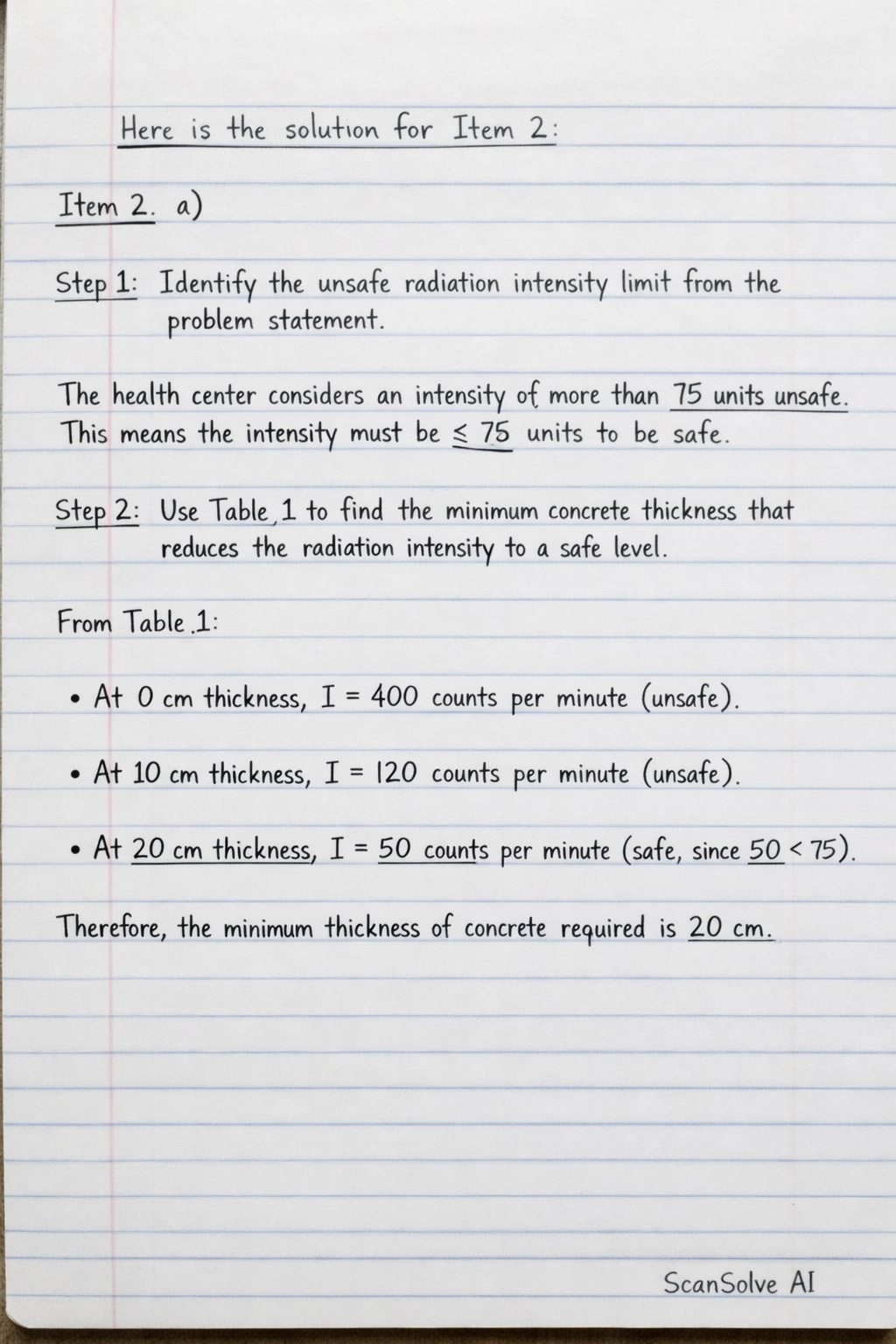
Identify the unsafe radiation intensity limit from the problem statement.
The health center considers an intensity of more than 75 units unsafe. This means the intensity must be 75 units or less to be safe.
Use Table 1 to find the minimum concrete thickness that reduces the radiation intensity to a safe level.
From Table 1:
- At thickness, intensity is (unsafe).
- At thickness, intensity is (unsafe).
- At thickness, intensity is (safe, as ). Therefore, the minimum thickness of concrete required is .
Explain why a specially constructed room is needed.
A specially constructed room with thick concrete walls is required to provide adequate shielding against the ionizing radiation emitted by the radioactive source. This shielding reduces the radiation intensity to safe levels, protecting health workers, patients, and the public from harmful exposure.
Part b
- Irradiation is the process of exposing an object or person to radiation from an external source. The object itself does not become radioactive.
- Contamination occurs when radioactive material is deposited on or inside an object or person, making the object or person radioactive.
Two precautions health workers should observe when handling the source:
- Minimize exposure time: Spend as little time as possible near the source to reduce the total radiation dose received.
- Use shielding: Utilize lead aprons, lead screens, or other appropriate barriers between themselves and the source to absorb radiation.
- Maintain distance: Keep as far away from the source as practically possible, as radiation intensity decreases significantly with distance.
- Wear protective clothing: Use gloves, lab coats, and other personal protective equipment to prevent direct contact and contamination.
Part c
Radioactive materials are useful in the treatment and diagnosis of diseases:
- Diagnosis: Radioactive tracers (e.g., Technetium-99m) are introduced into the body. Their movement and accumulation can be tracked using detectors to image organs, identify tumors, or detect blockages (e.g., in PET scans or SPECT scans).
- Treatment: In radiotherapy, high-energy radiation (e.g., from Cobalt-60) is directed at cancerous cells to damage their DNA and destroy them, preventing their growth and spread.
Part d
The radiation used to obtain an image of a patient's bone fracture is typically X-rays.
- Image production: X-rays are passed through the patient's body. Bones are denser than soft tissues, so they absorb more X-rays. Less radiation passes through the bones to reach the detector (film or digital sensor), creating a lighter image of the bones. Soft tissues absorb less radiation, appearing darker. A fracture appears as a break or discontinuity in the bone's structure.
One other application of this type of radiation (X-rays) is:
- Industrial radiography: Used to inspect welds, castings, and other materials for flaws or cracks without damaging the material.
- Security screening: Used in airports to scan luggage for prohibited items.
2 zimekamilika. Zimebaki 3 leo — tuma inayofuata.
Still stuck on this one?
Ask a follow-up, and the answer starts from this question.
Handwritten Step-by-Step Solution
Get this as a handwritten image on WhatsApp — like a tutor wrote it for you.
Try on WhatsApp