
This biology question covers important biological concepts and processes. The step-by-step explanation below helps you understand the underlying mechanisms and reasoning.
ScanSolve AI Answer
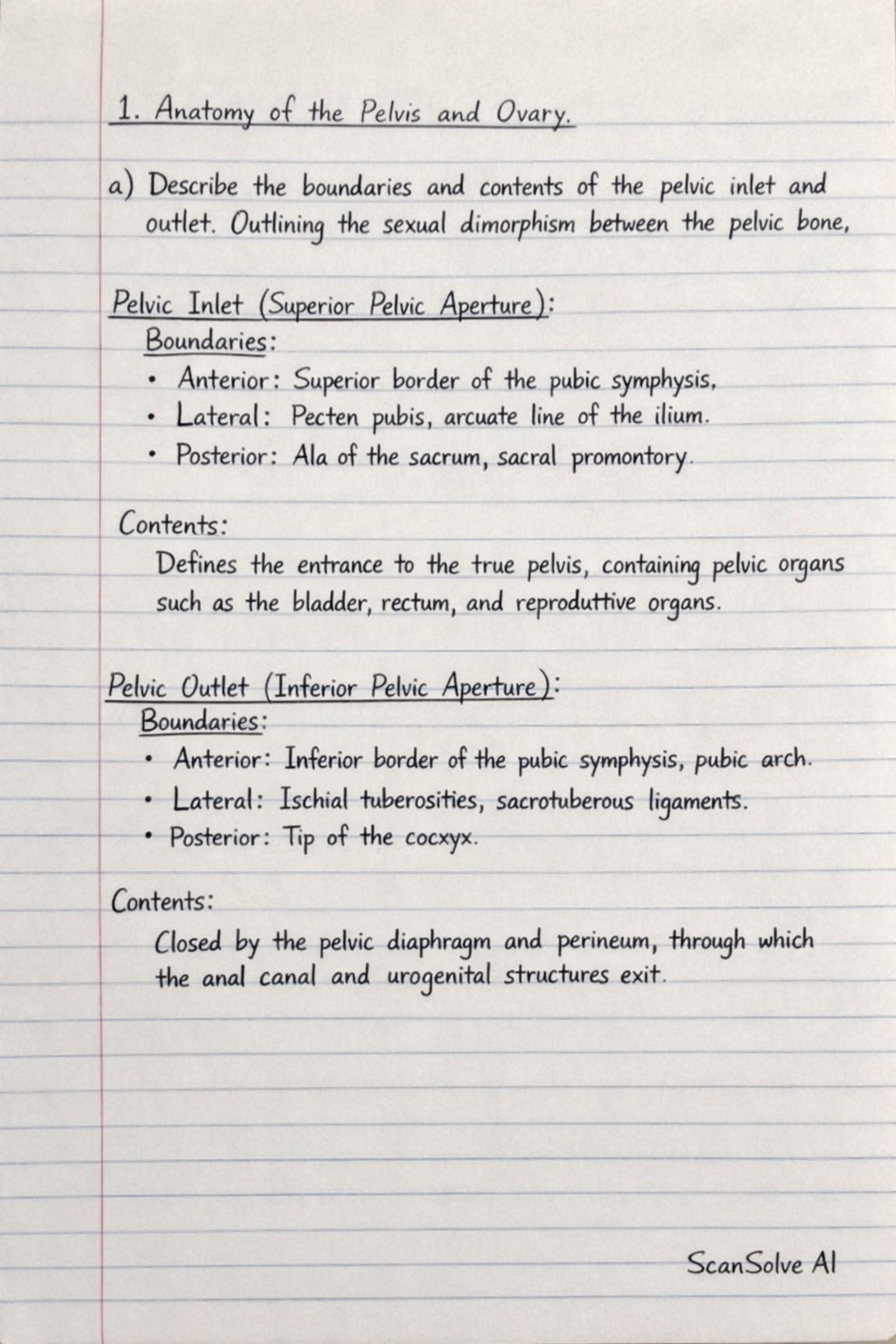
Here are the answers to the questions in an exam style: 1. Anatomy of the Pelvis and Ovary a) Describe the boundaries and contents of the pelvic inlet and outlet. Outlining the sexual dimorphism between the pelvic bone. Pelvic Inlet (Superior Pelvic Aperture): Boundaries: Anterior: Superior border of the pubic symphysis. Lateral: Pecten pubis, arcuate line of the ilium. Posterior: Ala of the sacrum, sacral promontory. Contents: It defines the entrance to the true pelvis, containing pelvic organs such as the bladder, rectum, and reproductive organs. Pelvic Outlet (Inferior Pelvic Aperture): Boundaries: Anterior: Inferior border of the pubic symphysis, pubic arch. Lateral: Ischial tuberosities, sacrotuberous ligaments. Posterior: Tip of the coccyx. Contents: It is closed by the pelvic diaphragm and perineum, through which the anal canal and urogenital structures exit. Sexual Dimorphism of the Pelvic Bone: Female Pelvis: General Structure:* Lighter, thinner, smoother. Pelvic Inlet:* Wider, more oval or rounded. Pelvic Outlet:* Wider, larger. Subpubic Angle (Pubic Arch):* Wider, usually > 80^ to 90^. Greater Sciatic Notch:* Wider. Ischial Tuberosities:* Everted. Sacrum:* Shorter, wider, less curved. Acetabulum:* Smaller, faces more anteriorly. Male Pelvis: General Structure:* Heavier, thicker, rougher, with more prominent muscle attachments. Pelvic Inlet:* Narrower, heart-shaped. Pelvic Outlet:* Narrower, smaller. Subpubic Angle (Pubic Arch):* Narrower, usually < 70^. Greater Sciatic Notch:* Narrower. Ischial Tuberosities:* Inverted. Sacrum:* Longer, narrower, more curved. Acetabulum:* Larger, faces more laterally. b) Discuss the gross Anatomy of the ovary. Add notes on its clinical correlations. Gross Anatomy of the Ovary: Description: Paired, almond-shaped female gonads, typically whitish-grey. Size: Approximately 3 cm × 1.5 cm × 1 cm in nulliparous women, decreasing in size after menopause. Location: Located in the ovarian fossa on the lateral pelvic wall, posterior to the broad ligament and anterior to the ureter and internal iliac artery. Surfaces: Medial and lateral. Borders: Anterior (mesovarian) and posterior (free). Poles: Superior (tubal) and inferior (uterine). Attachments: Mesovarium:* A fold of peritoneum, part of the broad ligament, attaching the anterior border of the ovary to the posterior surface of the broad ligament. Suspensory Ligament of the Ovary (Infundibulopelvic Ligament):* Extends from the tubal pole to the lateral pelvic wall, containing the ovarian artery, vein, lymphatics, and nerves. Ovarian Ligament:* Connects the uterine pole of the ovary to the lateral aspect of the uterus, inferior to the uterine tube. Blood Supply: Arterial:* Ovarian artery (direct branch of the abdominal aorta). Venous:* Ovarian veins (right drains into IVC, left drains into left renal vein). Lymphatic Drainage: Para-aortic lymph nodes (at the level of L1). Nerve Supply: Autonomic nerves from the ovarian plexus (T10-T11 spinal segments). Clinical Correlations: Ovarian Cysts: Fluid-filled sacs within or on the surface of an ovary, common and often benign, but can cause pain or rupture. Ovarian Torsion: Twisting of the ovary and its vascular pedicle, leading to ischemia and severe pain, requiring emergency surgery. Ectopic Pregnancy: Rarely, a fertilized egg can implant on the surface of the ovary, leading to an ovarian ectopic pregnancy. Polycystic Ovary Syndrome (PCOS): A hormonal disorder causing enlarged ovaries with small cysts on the outer edges, leading to irregular periods, excess androgen, and infertility. Ovarian Cancer: Often asymptomatic in early stages, making early detection difficult. Can spread rapidly. 2. Mediastinum Describe the mediastinum under the following: a) Anatomical boundaries The mediastinum is the central compartment of the thoracic cavity, located between the two pleural sacs. Superior Boundary: Thoracic inlet (superior aperture of the thorax). Inferior Boundary: Diaphragm. Anterior Boundary: Sternum. Posterior Boundary: Thoracic vertebral bodies (T1-T12). Lateral Boundaries: Mediastinal pleura of the lungs. The mediastinum is further subdivided by an imaginary transverse plane passing from the sternal angle (T4/T5 intervertebral disc) into: Superior Mediastinum: Above the transverse plane. Inferior Mediastinum: Below the transverse plane, which is further divided into: Anterior Mediastinum: Between the sternum and pericardium. Middle Mediastinum: Contains the pericardium and heart. Posterior Mediastinum: Between the pericardium and vertebral bodies. b) Contents Superior Mediastinum: Muscles: Origins of sternohyoid and sternothyroid muscles. Arteries: Arch of aorta and its three major branches (brachiocephalic trunk, left common carotid artery, left subclavian artery). Veins: Superior vena cava (SVC) and its tributaries (brachiocephalic veins), azygos vein (terminal part). Nerves: Vagus nerves (CN X), phrenic nerves, cardiac nerves, left recurrent laryngeal nerve. Lymphatics: Thoracic duct, lymph nodes. Other: Trachea, esophagus, thymus (in children). Inferior Mediastinum: Anterior Mediastinum: Thymus (remnant in adults). Loose connective tissue, fat. Lymph nodes. Internal thoracic vessels (branches). Middle Mediastinum: Heart and pericardium. Roots of great vessels (ascending aorta, pulmonary trunk, SVC, IVC, pulmonary veins). Phrenic nerves. Main bronchi. Lymph nodes. Posterior Mediastinum: Arteries: Descending thoracic aorta and its branches. Veins: Azygos, hemiazygos, and accessory hemiazygos veins. Nerves: Vagus nerves (esophageal plexus), splanchnic nerves (greater, lesser, least). Lymphatics: Thoracic duct, posterior mediastinal lymph nodes. Other: Esophagus. c) Clinical importance Mediastinitis: Inflammation of the mediastinum, often a severe infection resulting from esophageal perforation or spread from adjacent structures. Can be acute or chronic. Mediastinal Tumors: Can arise from any of the mediastinal contents (e.g., thymomas, lymphomas, neurogenic tumors, germ cell tumors). Symptoms depend on the size and location, often due to compression of surrounding structures (e.g., SVC syndrome, dysphagia, hoarseness). Mediastinal Shift: Displacement of mediastinal structures (heart, trachea) due to conditions like pneumothorax, hemothorax, or large pleural effusions, which can compromise cardiopulmonary function. Aortic Aneurysms: Dilation of the aorta within the mediastinum can compress adjacent structures or rupture, leading to life-threatening hemorrhage. Tracheal Compression: Enlarged lymph nodes or tumors can compress the trachea, causing dyspnea or stridor. Esophageal Disorders: The esophagus's passage through the posterior mediastinum makes it susceptible to disorders like achalasia, strictures, or esophageal cancer, which can present with dysphagia. 3. Short Notes a) Dural venous sinuses The dural venous sinuses are endothelium-lined spaces between the two layers of the dura mater (endosteal and meningeal layers) of the brain. They are valveless channels that collect venous blood from the brain, cranial bones, and meninges, ultimately draining into the internal jugular veins. Major sinuses include the superior sagittal, inferior sagittal, straight, transverse, sigmoid, cavernous, and petrosal sinuses. They are crucial for cerebral venous drainage and cerebrospinal fluid reabsorption (via arachnoid granulations). Clinically, they are susceptible to thrombosis (e.g., cavernous sinus thrombosis), which can lead to increased intracranial pressure, neurological deficits, and can be life-threatening due to their connections with facial veins. b) Brown-Sequard syndrome Brown-Sequard syndrome is a rare neurological condition caused by a hemicord lesion (damage to one half of the spinal cord). This typically results from trauma (e.g., stab wound), tumor, or ischemia. The syndrome presents with a characteristic pattern of neurological deficits below the level of the lesion: Ipsilateral (same side as lesion): Loss of voluntary motor function* (corticospinal tract damage). Loss of proprioception and vibratory sensation* (dorsal column damage). Contralateral (opposite side of lesion): Loss of pain and temperature sensation* (spinothalamic tract damage), typically starting 1-2 segments below the lesion due to decussation. This distinct presentation helps in localizing spinal cord injuries. c) Visual pathway The visual pathway is the neurological route by which visual information travels from the retina to the visual cortex of the brain. 1. Retina: Photoreceptors (rods and cones) convert light into electrical signals. Ganglion cells in the retina form the optic nerve. 2. Optic Nerve (CN II): Carries signals from each eye. 3. Optic Chiasm: Located anterior to the pituitary gland, where fibers from the nasal (medial) half of each retina decussate (cross over) to the contralateral side. Temporal (lateral) fibers remain ipsilateral. 4. Optic Tracts: After the chiasm, each optic tract contains fibers from the temporal half of the ipsilateral retina and the nasal half of the contralateral retina, thus carrying information from the opposite visual field. 5. Lateral Geniculate Nucleus (LGN) of Thalamus: Most fibers synapse here. The LGN processes and relays visual information. 6. Optic Radiations (Geniculocalcarine Tract): Fibers from the LGN project through the parietal and temporal lobes (Meyer's loop) to the primary visual cortex. 7. Primary Visual Cortex (Brodmann Area 17): Located in the occipital lobe, where visual information is initially processed and perceived. Damage at different points along this pathway results in specific visual field defects (e.g., hemianopia, quadrantanopia). d) Microscopic anatomy of the male gonad The male gonad is the testis, an oval-shaped organ responsible for spermatogenesis and hormone production. Tunics: Tunica Vaginalis:* Outer serous sac, derived from peritoneum, with parietal and visceral layers. Tunica Albuginea: Dense, fibrous capsule beneath the tunica vaginalis, which thickens posteriorly to form the mediastinum testis*. Tunica Vasculosa:* Inner vascular layer lining the septa and covering the inner surface of the tunica albuginea. Seminiferous Tubules: Coiled tubules within lobules (separated by septa extending from the mediastinum testis) where spermatogenesis* occurs. Lined by germinal epithelium* containing: Spermatogenic cells:* Undergo meiosis to produce spermatozoa. Sertoli cells (sustentacular cells):* Support, nourish, and protect developing sperm; form the blood-testis barrier; produce inhibin and androgen-binding protein. Interstitial Tissue: Connective tissue between the seminiferous