
We read your photo as:
Draw a diagram of the female pelvic organs and describe the vagina in terms of its relations, size, epithelial lining, and antimicrobial adaptations.
ScanSolve AI Answer
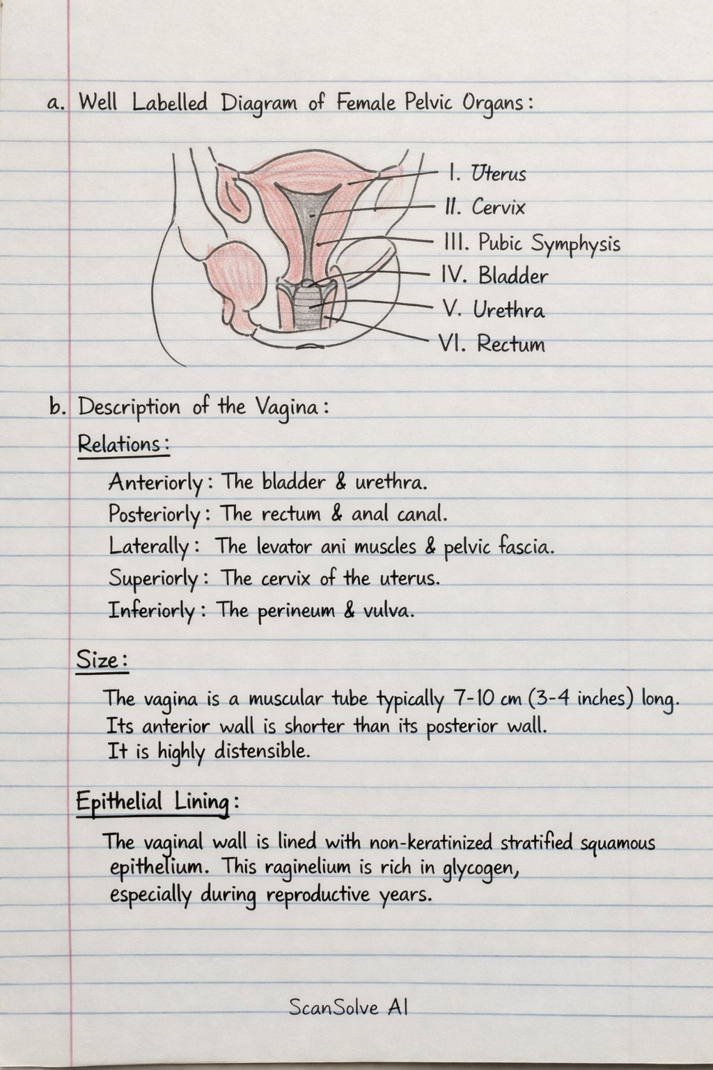
a. Draw a well labelled diagram of the structure below
As an AI, I cannot draw. However, I can identify the parts labeled in the provided diagram of the female pelvic organs.
- I: Uterus
- II: Cervix
- III: Pubic symphysis
- IV: Bladder
- V: Urethra
- VI: Rectum
b. Describe the vagina in terms of:
-
Relations
- Anteriorly: The bladder and urethra.
- Posteriorly: The rectum and anal canal.
- Laterally: The levator ani muscles and pelvic fascia.
- Superiorly: The cervix of the uterus.
- Inferiorly: The perineum and vulva.
-
Size The vagina is a muscular tube typically 7-10 cm (3-4 inches) long. Its anterior wall is usually shorter than its posterior wall. It is highly distensible.
-
Epithelial lining The vaginal wall is lined with non-keratinized stratified squamous epithelium. This epithelium is rich in glycogen, especially during reproductive years.
-
Antimicrobial adaptations
- Acidic pH: The normal vaginal environment is acidic (pH 3.8-4.5) due to the production of lactic acid by Lactobacillus bacteria, which ferment glycogen from the epithelial cells. This acidity inhibits the growth of many pathogens.
- Normal flora: A healthy population of beneficial bacteria, primarily Lactobacillus species, outcompetes and suppresses the growth of harmful microorganisms.
- Mucus production: Cervical glands produce mucus that helps to flush out pathogens and provides a physical barrier.
- Epithelial shedding: The constant shedding of the stratified squamous epithelial cells helps to remove attached pathogens.
Still stuck on this one?
Ask a follow-up, and the answer starts from this question.
More Biology Questions
Handwritten Step-by-Step Solution
Get this as a handwritten image on WhatsApp — like a tutor wrote it for you.
Try on WhatsApp