
We read your photo as:
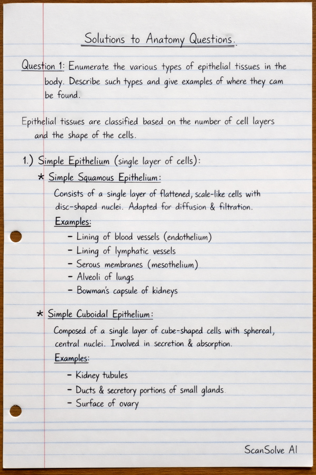
Enumerate the various types of epithelial tissues in the body. Describe each type and give examples of where they can be found.
ScanSolve AI Answer
Here are the solutions to your anatomy questions.
Question 1: Enumerate the various types of epithelial tissues in the body. Describe such type and give examples of where they can be found.
Epithelial tissues are classified based on the number of cell layers and the shape of the cells.
-
Simple Epithelium (single layer of cells):
- Simple Squamous Epithelium: Consists of a single layer of flattened, scale-like cells with disc-shaped nuclei. It is adapted for diffusion and filtration.
- Examples: Lining of blood vessels (endothelium), lining of lymphatic vessels, serous membranes (mesothelium), alveoli of lungs, Bowman's capsule of kidneys.
- Simple Cuboidal Epithelium: Composed of a single layer of cube-shaped cells with spherical, centrally located nuclei. It is involved in secretion and absorption.
- Examples: Kidney tubules, ducts and secretory portions of small glands, surface of ovary.
- Simple Columnar Epithelium: Made of a single layer of tall, column-shaped cells with oval nuclei usually located near the base. It functions in absorption and secretion. Some types have cilia or microvilli.
- Examples: Non-ciliated type lines most of the digestive tract (stomach to rectum), gallbladder. Ciliated type lines small bronchi, uterine tubes, and some regions of the uterus.
- Pseudostratified Columnar Epithelium: Appears stratified because nuclei are at different levels, but all cells rest on the basement membrane. Some cells are shorter and do not reach the apical surface. Often ciliated and contains goblet cells. Functions in secretion and propulsion of mucus.
- Examples: Non-ciliated type in male's sperm-carrying ducts and ducts of large glands. Ciliated type lines the trachea and most of the upper respiratory tract.
- Simple Squamous Epithelium: Consists of a single layer of flattened, scale-like cells with disc-shaped nuclei. It is adapted for diffusion and filtration.
-
Stratified Epithelium (two or more layers of cells):
- Stratified Squamous Epithelium: The most widespread stratified epithelium. The superficial layers are flattened squamous cells, while deeper layers are cuboidal or columnar. It protects underlying tissues in areas subject to abrasion.
- Examples: Keratinized type forms the epidermis of the skin. Non-keratinized type lines the esophagus, mouth, and vagina.
- Stratified Cuboidal Epithelium: Generally two layers of cuboidal cells. It functions in protection.
- Examples: Ducts of large glands (e.g., sweat glands, mammary glands, salivary glands).
- Stratified Columnar Epithelium: Several layers of cells; superficial cells are columnar, while basal cells are usually cuboidal. It functions in protection and secretion.
- Examples: Rare in the body; found in small amounts in the male urethra and in large ducts of some glands.
- Transitional Epithelium: Resembles both stratified squamous and stratified cuboidal. The basal cells are cuboidal or columnar, and the superficial cells are dome-shaped or squamous-like, depending on the degree of organ stretch. It allows for stretching.
- Examples: Lines the ureters, bladder, and part of the urethra.
- Stratified Squamous Epithelium: The most widespread stratified epithelium. The superficial layers are flattened squamous cells, while deeper layers are cuboidal or columnar. It protects underlying tissues in areas subject to abrasion.
Question 2: With the aid of a diagram describe the arterial circle of Willis. Add notes on its clinical importance.
The Arterial Circle of Willis is an anastomotic ring of arteries located at the base of the brain, surrounding the optic chiasm and the pituitary stalk. It provides a critical collateral circulation pathway to the brain, ensuring continuous blood supply even if one of the major arteries supplying the brain is partially or completely occluded.
Description of the Arterial Circle of Willis:
The Circle of Willis is formed by the following arteries:
- Posterior Cerebral Arteries (PCAs): These are the terminal branches of the basilar artery.
- Posterior Communicating Arteries (PCoAs): These connect the posterior cerebral arteries to the internal carotid arteries.
- Internal Carotid Arteries (ICAs): These enter the cranial cavity and give off the middle cerebral arteries (MCAs) and anterior cerebral arteries (ACAs).
- Anterior Cerebral Arteries (ACAs): These are branches of the internal carotid arteries.
- Anterior Communicating Artery (ACoA): This single artery connects the two anterior cerebral arteries, completing the anterior part of the circle.
The major arteries contributing to the circle are:
- Vertebral Arteries: These ascend through the neck, merge to form the basilar artery.
- Basilar Artery: This ascends along the brainstem and bifurcates into the two posterior cerebral arteries.
- Internal Carotid Arteries: These ascend through the neck, enter the skull, and each divides into the anterior cerebral artery and middle cerebral artery.
The arrangement is as follows: The two ACAs are connected anteriorly by the ACoA. The ACAs then connect to the ICAs. The ICAs give off the PCoAs, which connect to the PCAs. The PCAs are branches of the basilar artery, which is formed by the union of the vertebral arteries.
Clinical Importance:
The Circle of Willis is clinically significant due to its role in maintaining cerebral perfusion:
- Collateral Circulation: It acts as a safety mechanism. If one of the major arteries supplying the brain (e.g., internal carotid or vertebral artery) becomes narrowed or blocked, blood can be rerouted through the circle to supply the affected area, potentially preventing or minimizing ischemic damage (stroke).
- Aneurysms: The junctions of the arteries forming the Circle of Willis are common sites for the formation of saccular (berry) aneurysms. These are weak spots in the arterial wall that can balloon out and rupture, leading to a subarachnoid hemorrhage, a life-threatening condition. The most common sites for aneurysms are the anterior communicating artery, posterior communicating artery, and the trifurcation of the middle cerebral artery.
- Variations: There are significant anatomical variations in the completeness and size of the arteries forming the Circle of Willis among individuals. A "complete" circle is present in only about 20-25% of the population. Incomplete or hypoplastic segments can compromise its collateral function, making individuals more vulnerable to ischemic events.
- Ischemic Stroke: Understanding the Circle of Willis is crucial in diagnosing and managing ischemic strokes. The location of an occlusion relative to the circle determines the extent of collateral flow and the potential for neurological deficits.
Question 3:
a) Describe the gross anatomy of the thoracic inlet and outlet.
Thoracic Inlet (Superior Thoracic Aperture):
The thoracic inlet is the superior opening of the thoracic cavity, connecting it with the neck. It is kidney-shaped and slopes anteriorly and inferiorly.
- Boundaries:
- Anteriorly: Superior border of the manubrium of the sternum.
- Posteriorly: Anterior surface of the body of the first thoracic vertebra (T1).
- Laterally: Medial borders of the first ribs and their costal cartilages.
- Contents: Structures passing between the neck and thorax include:
- Trachea
- Esophagus
- Major blood vessels: Brachiocephalic veins, subclavian arteries, common carotid arteries (or brachiocephalic artery on the right), internal jugular veins.
- Nerves: Vagus nerves, phrenic nerves, recurrent laryngeal nerves, sympathetic trunks, brachial plexus.
- Apex of the lungs and pleura
- Lymphatic ducts: Thoracic duct (left), right lymphatic duct (right).
- Muscles: Sternohyoid, sternothyroid.
Thoracic Outlet (Inferior Thoracic Aperture):
The thoracic outlet is the inferior opening of the thoracic cavity, much larger than the inlet, and is largely closed by the diaphragm.
- Boundaries:
- Anteriorly: Xiphisternal joint.
- Anterolaterally: Costal cartilages of ribs 7-10, forming the costal margin.
- Posterolaterally: Ribs 11 and 12.
- Posteriorly: Body of the 12th thoracic vertebra (T12).
- Contents: The diaphragm largely closes this aperture, with several openings for structures passing between the thorax and abdomen:
- Aortic hiatus: For the aorta, thoracic duct, and azygos vein.
- Esophageal hiatus: For the esophagus and vagus nerves.
- Caval hiatus: For the inferior vena cava and right phrenic nerve.
- Other structures include sympathetic trunks, splanchnic nerves, and subcostal nerves and vessels.
b) Describe the gross anatomy of the mediastinum. Add notes on the clinical significance.
The mediastinum is the central compartment of the thoracic cavity, located between the two pleural sacs (lungs). It extends from the superior thoracic aperture to the diaphragm inferiorly, and from the sternum anteriorly to the vertebral column posteriorly.
Divisions of the Mediastinum:
A horizontal plane passing from the sternal angle (T4/T5 intervertebral disc) divides the mediastinum into:
- Superior Mediastinum: Located above the sternal angle.
- Contents: Thymus, great vessels (arch of aorta and its branches, superior vena cava, brachiocephalic veins), trachea, esophagus, thoracic duct, vagus nerves, phrenic nerves, recurrent laryngeal nerves, sympathetic trunks.
- Inferior Mediastinum: Located below the sternal angle. This is further subdivided by the pericardium (containing the heart) into:
- Anterior Mediastinum: Smallest part, anterior to the pericardium.
- Contents: Thymus remnants, lymph nodes, loose connective tissue.
- Middle Mediastinum: Contains the heart and pericardium.
- Contents: Heart, pericardium, roots of great vessels (ascending aorta, pulmonary trunk, superior and inferior vena cava), phrenic nerves, main bronchi.
- Posterior Mediastinum: Posterior to the pericardium.
- Contents: Esophagus, descending thoracic aorta, azygos and hemiazygos veins, thoracic duct, vagus nerves, sympathetic trunks, posterior mediastinal lymph nodes.
- Anterior Mediastinum: Smallest part, anterior to the pericardium.
Clinical Significance:
- Mediastinal Shift: Displacement of the mediastinum from its normal central position, often due to conditions like pneumothorax, hemothorax, or large pleural effusions. This can compress the great vessels and heart, leading to cardiovascular compromise.
- Mediastinal Masses/Tumors: The mediastinum is a common site for tumors (e.g., thymomas, lymphomas, neurogenic tumors, germ cell tumors, thyroid masses). Symptoms depend on the location and structures compressed (e.g., superior vena cava syndrome, dysphagia, hoarseness).
- Mediastinitis: Inflammation of the mediastinum, often a severe infection resulting from esophageal perforation or spread from adjacent structures. It is a life-threatening condition.
- Aortic Aneurysms: Aneurysms of the thoracic aorta can expand into the mediastinum, compressing surrounding structures or rupturing, leading to catastrophic hemorrhage.
- Tracheal Compression: Enlarged lymph nodes or tumors in the mediastinum can compress the trachea, causing respiratory distress.
Question 4:
a) Explain the anatomy of the perineal pouch.
The perineal pouch refers to two distinct fascial-lined spaces within the perineum: the superficial perineal pouch and the deep perineal pouch. The perineum is the diamond-shaped region inferior to the pelvic diaphragm, between the thighs.
1. Superficial Perineal Pouch:
- Location: A potential space inferior to the perineal membrane and superior to the superficial perineal fascia (Colles' fascia).
- Boundaries:
- Inferiorly: Superficial perineal fascia (Colles' fascia).
- Superiorly: Perineal membrane.
- Laterally: Ischiopubic rami.
- Posteriorly: Fused superficial perineal fascia and perineal membrane.
- Anteriorly: Open to the anterior abdominal wall.
- Contents (Male):
- Root of the penis (bulb of penis and crura of penis).
- Ischiocavernosus muscles (covering the crura).
- Bulbospongiosus muscles (covering the bulb).
- Spongy urethra (proximal part).
- Superficial transverse perineal muscles.
- Perineal branches of pudendal nerves and internal pudendal vessels.
- Contents (Female):
- Clitoris (crura and body).
- Ischiocavernosus muscles.
- Bulbospongiosus muscles (covering the bulbs of the vestibule).
- Bulbs of the vestibule.
- Greater vestibular glands (Bartholin's glands).
- Superficial transverse perineal muscles.
- Perineal branches of pudendal nerves and internal pudendal vessels.
2. Deep Perineal Pouch:
- Location: A space superior to the perineal membrane and inferior to the pelvic diaphragm.
- Boundaries:
- Inferiorly: Perineal membrane.
- Superiorly: Inferior fascia of the pelvic diaphragm.
- Laterally: Obturator internus fascia.
- Contents (Male):
- External urethral sphincter.
- Deep transverse perineal muscles.
- Bulbourethral glands (Cowper's glands).
- Proximal part of the membranous urethra.
- Dorsal neurovasculature of the penis.
- Contents (Female):
- External urethral sphincter.
- Deep transverse perineal muscles.
- Proximal part of the urethra.
- Proximal part of the vagina.
- Dorsal neurovasculature of the clitoris.
b) Give an account of the gross anatomy the uterus, add notes on the histology.
Gross Anatomy of the Uterus:
The uterus is a hollow, pear-shaped muscular organ located in the lesser pelvis, between the urinary bladder anteriorly and the rectum posteriorly. It is the primary female reproductive organ responsible for housing and nourishing a developing fetus.
- Size and Shape: Approximately 7.5 cm long, 5 cm wide, and 2.5 cm thick in nulliparous women. It is larger in multiparous women.
- Position: Typically anteverted (tilted forward at the junction of the cervix and vagina) and anteflexed (flexed forward at the junction of the body and cervix). Its position is variable depending on bladder and rectal fullness.
- Parts:
- Fundus: The rounded, superior part of the uterus, superior to the entrance of the uterine
Still stuck on this one?
Ask a follow-up, and the answer starts from this question.
More Biology Questions
Handwritten Step-by-Step Solution
Get this as a handwritten image on WhatsApp — like a tutor wrote it for you.
Try on WhatsApp