
We read your photo as:
Describe the formation, relations, and branches of the brachial plexus.
Biology
ScanSolve AI Answer
1)
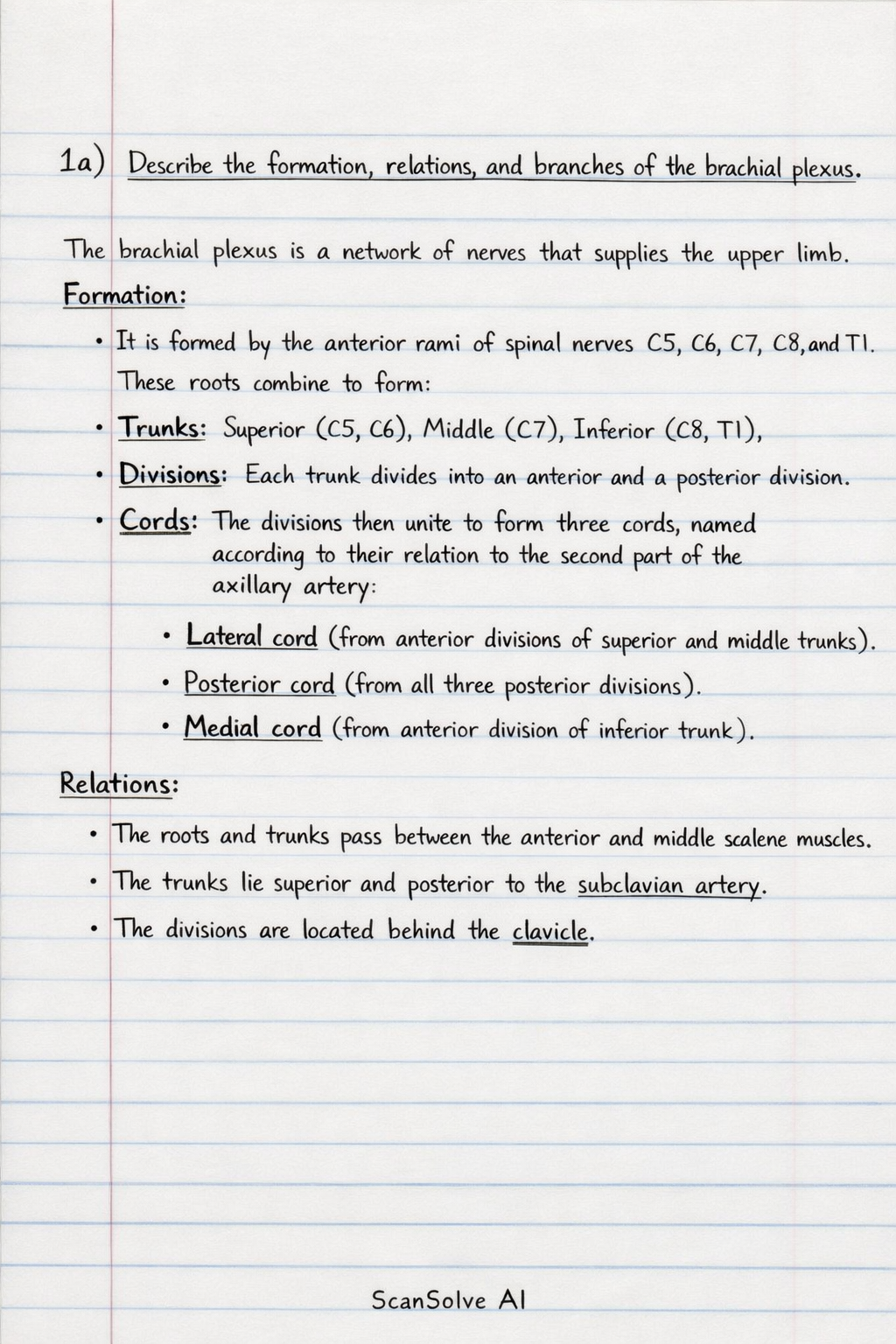
a) Describe the formation, relations, and branches of the brachial plexus.
The brachial plexus is a network of nerves that supplies the upper limb.
- Formation: It is formed by the anterior rami of spinal nerves C5, C6, C7, C8, and T1. These roots combine to form:
- Trunks: Superior (C5, C6), Middle (C7), Inferior (C8, T1).
- Divisions: Each trunk divides into an anterior and a posterior division.
- Cords: The divisions then unite to form three cords, named according to their relation to the second part of the axillary artery:
- Lateral cord (from anterior divisions of superior and middle trunks).
- Posterior cord (from all three posterior divisions).
- Medial cord (from anterior division of inferior trunk).
- Relations:
- The roots and trunks pass between the anterior and middle scalene muscles.
- The trunks lie superior and posterior to the subclavian artery.
- The divisions are located behind the clavicle.
- The cords are closely related to the axillary artery in the axilla, with the lateral cord lateral to it, the posterior cord posterior to it, and the medial cord medial to it.
- Branches:
- From Roots: Dorsal scapular nerve (C5), Long thoracic nerve (C5, C6, C7).
- From Trunks: Suprascapular nerve (superior trunk), Nerve to subclavius (superior trunk).
- From Cords:
- Lateral Cord: Lateral pectoral nerve, Musculocutaneous nerve, Lateral root of median nerve.
- Posterior Cord: Upper subscapular nerve, Thoracodorsal nerve, Lower subscapular nerve, Axillary nerve, Radial nerve.
- Medial Cord: Medial pectoral nerve, Medial cutaneous nerve of arm, Medial cutaneous nerve of forearm, Ulnar nerve, Medial root of median nerve.
b) Name this condition and discuss its anatomical basis, nerve roots involved and the muscles affected.
- Name of condition: Erb's Palsy (or Erb-Duchenne palsy).
- Anatomical Basis: This condition results from injury to the upper trunk of the brachial plexus, typically due to excessive traction or stretching of the neck and shoulder during a difficult delivery (shoulder dystocia). This causes damage to the C5 and C6 nerve roots.
- Nerve Roots Involved: C5 and C6.
- Muscles Affected: The injury to C5 and C6 affects nerves derived from the upper trunk, leading to paralysis or weakness in muscles responsible for shoulder abduction, lateral rotation, and elbow flexion.
- Deltoid and Supraspinatus (supplied by axillary and suprascapular nerves) – impaired shoulder abduction.
- Infraspinatus and Teres minor (supplied by suprascapular and axillary nerves) – impaired lateral rotation of the shoulder.
- Biceps brachii and Brachialis (supplied by musculocutaneous nerve) – impaired elbow flexion and forearm supination.
- Brachioradialis (supplied by radial nerve) – weakened elbow flexion. The characteristic "waiter's tip" position (arm adducted, medially rotated, and elbow extended) results from the unopposed action of unaffected muscles and the paralysis of the affected muscles.
2) Discuss the axilla under the following headings; (i) Boundaries (ii) Contents (iii) Relations and (iv) Clinical correlates.
The axilla is a pyramidal space between the upper part of the arm and the side of the chest.
-
(i) Boundaries:
- Apex: Formed by the cervicoaxillary canal, bounded by the clavicle anteriorly, the superior border of the scapula posteriorly, and the first rib medially.
- Base: Formed by the skin and axillary fascia, extending from the arm to the thoracic wall.
- Anterior Wall: Pectoralis major, pectoralis minor, and the clavipectoral fascia.
- Posterior Wall: Subscapularis, teres major, and latissimus dorsi muscles.
- Medial Wall: Upper four or five ribs and the intercostal muscles covered by the serratus anterior muscle.
- Lateral Wall: Intertubercular groove of the humerus.
-
(ii) Contents:
- Axillary artery and its branches.
- Axillary vein and its tributaries.
- Brachial plexus (cords and terminal branches).
- Axillary lymph nodes and associated lymphatic vessels.
- Long thoracic nerve and intercostobrachial nerve.
- Axillary fat and areolar tissue.
-
(iii) Relations:
- Superiorly: Continuous with the root of the neck via the cervicoaxillary canal.
- Inferiorly: Continuous with the arm and the superficial fascia of the chest.
- Anteriorly: Related to the pectoral region.
- Posteriorly: Related to the scapular region.
- Medially: Related to the lateral thoracic wall.
- Laterally: Related to the proximal part of the humerus.
-
(iv) Clinical Correlates:
- Axillary Lymphadenopathy: Swelling of axillary lymph nodes, often indicating infection in the upper limb or breast, or metastasis of breast cancer.
- Axillary Nerve Injury: Can occur due to shoulder dislocation or fracture of the surgical neck of the humerus, leading to paralysis of the deltoid and teres minor muscles, resulting in loss of shoulder abduction and a flattened shoulder contour.
- Brachial Plexus Injuries: Trauma to the axilla can damage the brachial plexus, leading to motor and sensory deficits in the upper limb.
- Axillary Artery Compression: The axillary artery can be compressed against the humerus in cases of trauma or used for pulse palpation.
- Intercostobrachial Nerve Damage: This nerve, which supplies sensation to the medial arm, can be damaged during axillary surgery (e.g., mastectomy with axillary lymph node dissection), leading to numbness in the area.
Was this helpful?
Still stuck on this one?
Ask a follow-up, and the answer starts from this question.
Got a different question?Ask your own question
More Biology Questions
QSummarize the divisions and cords of the brachial plexus, including their main branches.QDoes the brachial artery lie lateral or medial to the median nerveQIdentify the branches of the axillary artery and the correct statement regarding its origin and…QWhich spinal nerve segments primarily mediate the brachioradialis reflex?QQ1: The median nerve is formed by the union of:
Handwritten Step-by-Step Solution
Get this as a handwritten image on WhatsApp — like a tutor wrote it for you.
Try on WhatsApp