
We read your photo as:
Describe the histological procedure for preparing permanent stained slides of testes and ovaries at different reproductive stages.
Biology
ScanSolve AI Answer
This study involves preparing permanent stained slides to examine the microscopic structure (histology) of testes and ovaries at different reproductive stages, correlating structural changes with their functions.
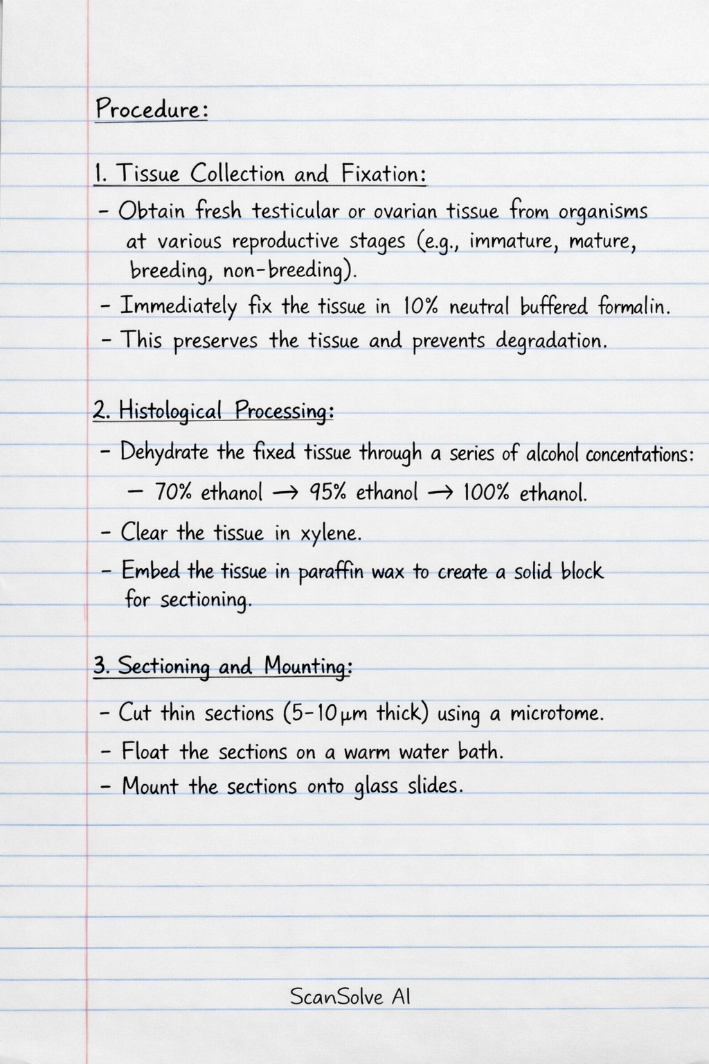
Procedure:
- Tissue Collection and Fixation: Obtain fresh testicular or ovarian tissue from organisms representing various reproductive phases (e.g., immature, mature, breeding, non-breeding). Immediately fix the tissue in a preservative like 10% neutral buffered formalin to prevent degradation and preserve cellular architecture.
- Histological Processing: Dehydrate the fixed tissue through a series of increasing alcohol concentrations, clear it in xylene, and then embed it in paraffin wax. This provides a solid block for sectioning.
- Sectioning and Mounting: Cut thin sections (typically 5-10 µm thick) from the paraffin block using a microtome. These sections are then floated on a warm water bath and mounted onto glass slides.
- Staining: Remove paraffin from the sections, rehydrate them, and then stain them using histological dyes. Hematoxylin and Eosin (H&E) is a common stain, where hematoxylin stains nuclei blue/purple and eosin stains cytoplasm and extracellular matrix pink.
- Permanent Mounting: Dehydrate the stained sections, clear them, and then mount a coverslip over them using a permanent mounting medium (e.g., DPX) to create a permanent slide.
- Microscopic Examination: Observe the prepared slides under a light microscope at various magnifications.
Key Functional Observations:
- Testes: Examine the seminiferous tubules for stages of spermatogenesis, the presence and activity of Sertoli cells (support cells), and Leydig cells (interstitial cells producing testosterone). Changes in these structures reflect sperm production and hormone synthesis across different reproductive phases.
- Ovaries: Observe the various stages of follicle development (primordial, primary, secondary, Graafian follicles), the presence of oocytes, and structures like the corpus luteum (after ovulation) or corpus albicans (regressed corpus luteum). These indicate cyclical changes in egg maturation and ovulation, as well as hormone production.
This study provides insights into the cellular and tissue-level adaptations of reproductive organs during different physiological states.
Was this helpful?
Still stuck on this one?
Ask a follow-up, and the answer starts from this question.
Got a different question?Ask your own question
More Biology Questions
QList four microtome knives and six basic steps in staining and mounting paraffin sections.QThe following are the common Romanowsky stain used in parasitology procedures, exceptQDescribe the processes involved in tissue processingQStages of preparing compound microscopQDescribe the microscopic anatomy of the male gonad, including its tunics, seminiferous tubules, and…
Handwritten Step-by-Step Solution
Get this as a handwritten image on WhatsApp — like a tutor wrote it for you.
Try on WhatsApp