
We read your photo as:
You are given a whole blood sample labeled A. With the materials provided, a) Determine ABO and Rhesus group of sample A. b) Determine the PCV of sample A and comment on your result.
ScanSolve AI Answer
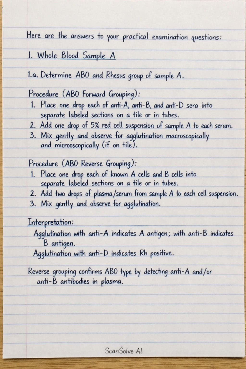
Here are the answers to your practical examination questions:
1. Whole Blood Sample A
-
1.a. Determine ABO and Rhesus group of sample A.
- Procedure (ABO Forward Grouping):
- Place one drop each of anti-A, anti-B, and anti-D sera into separate labeled sections on a tile or in tubes.
- Add one drop of a 5% red cell suspension of sample A to each serum.
- Mix gently and observe for agglutination macroscopically and microscopically (if on tile).
- Procedure (ABO Reverse Grouping):
- Place one drop each of known A cells and B cells into separate labeled sections on a tile or in tubes.
- Add two drops of plasma/serum from sample A to each cell suspension.
- Mix gently and observe for agglutination.
- Interpretation: Agglutination with anti-A indicates A antigen; with anti-B indicates B antigen. Agglutination with anti-D indicates Rh positive. Reverse grouping confirms ABO type by detecting anti-A and/or anti-B antibodies in plasma.
- Procedure (ABO Forward Grouping):
-
1.b. Determine the PCV of sample A and comment on your result.
- Procedure:
- Fill a plain capillary tube (for non-anticoagulated blood) or a heparinized capillary tube (for anticoagulated blood) with sample A, leaving about 15 mm unfilled.
- Seal one end of the capillary tube with clay or by heating.
- Place the sealed tube in a microhematocrit centrifuge with the sealed end facing outwards.
- Centrifuge at 10,000-12,000 rpm for 5 minutes.
- Read the Packed Cell Volume (PCV) using a microhematocrit reader.
- Comment on Result: Compare the obtained PCV value to the normal reference range (e.g., 0.40-0.54 L/L for adult males, 0.37-0.47 L/L for adult females). A value below the range suggests anemia, while a value above the range suggests polycythemia.
- Procedure:
-
1.c. Make a suitable film of sample A and stain for determination of red cell morphology.
- Procedure (Thin Film Preparation):
- Place a small drop of blood (sample A) near one end of a clean, grease-free glass slide.
- Place a spreader slide in front of the blood drop at a 30-45 degree angle.
- Draw the spreader back into the drop, allowing the blood to spread along its edge.
- Push the spreader smoothly and rapidly forward to create a thin film with a feathered edge.
- Allow the film to air dry completely.
- Procedure (Staining - e.g., Leishman stain):
- Place the dried film on a staining rack.
- Flood the slide with undiluted Leishman stain for 1-2 minutes (for fixation).
- Add an equal volume of buffered distilled water (pH 6.8) to the stain, mix gently by blowing, and allow to stand for 5-10 minutes (for staining).
- Wash gently with buffered distilled water until the film appears pinkish-purple.
- Allow to air dry vertically.
- Procedure (Thin Film Preparation):
-
1.d. What 2 precautions did you take when making the film.
- Ensure the glass slides are clean and grease-free to prevent artifacts and ensure even spreading of the blood.
- Use the correct angle and speed when pushing the spreader slide to produce a thin, uniform film with a good feathered edge, avoiding thick areas or streaks.
2. Histological Sections B and C
-
2.a. Stain slide B to demonstrate general tissue morphology.
- Procedure (Hematoxylin and Eosin - H&E stain):
- Deparaffinize slide B in xylene (2 changes, 5 minutes each).
- Rehydrate through graded alcohols (100%, 90%, 70% ethanol, 2 minutes each) to distilled water.
- Stain with Hematoxylin for 5-10 minutes.
- Wash in running tap water for 5 minutes.
- Differentiate in 1% acid alcohol (1-2 dips).
- Blue in Scott's tap water or 0.2% ammonia water for 1-2 minutes.
- Wash in running tap water for 5 minutes.
- Stain with Eosin for 1-5 minutes.
- Dehydrate through graded alcohols (70%, 90%, 100% ethanol, 2 minutes each).
- Clear in xylene (2 changes, 5 minutes each).
- Mount with a coverslip using a suitable mounting medium.
- Procedure (Hematoxylin and Eosin - H&E stain):
-
2.b. Stain slide C to demonstrate connective tissue.
- Procedure (Masson's Trichrome stain):
- Deparaffinize slide C in xylene (2 changes, 5 minutes each).
- Rehydrate through graded alcohols to distilled water.
- Mordant in Bouin's solution at for 1 hour or at room temperature overnight.
- Wash in running tap water until yellow color disappears (approx. 5-10 minutes).
- Stain with Weigert's Iron Hematoxylin for 10 minutes.
- Wash in running tap water for 5 minutes.
- Stain with Biebrich Scarlet-Acid Fuchsin solution for 10-15 minutes.
- Rinse in distilled water.
- Differentiate in Phosphomolybdic-Phosphotungstic Acid solution for 10-15 minutes.
- Stain with Aniline Blue solution for 5-10 minutes.
- Rinse in distilled water.
- Differentiate in 1% Acetic Acid solution for 2-5 minutes.
- Dehydrate rapidly through 95% and absolute alcohol.
- Clear in xylene (2 changes).
- Mount with a coverslip.
- Expected Result: Collagen fibers stain blue, nuclei stain black, cytoplasm, muscle, and red blood cells stain red.
- Procedure (Masson's Trichrome stain):
3. Anaerobic Culture Sample D
-
3.a. Make a film and stain by Gram's method. Comment on your findings.
- Procedure (Gram Stain):
- Prepare a thin smear of sample D on a clean glass slide and air dry.
- Heat fix the smear by passing it through a flame a few times.
- Flood the smear with Crystal Violet for 1 minute.
- Rinse gently with water.
- Flood with Gram's Iodine for 1 minute.
- Rinse gently with water.
- Decolorize with Acetone-Alcohol for 10-20 seconds until the runoff is clear.
- Rinse immediately with water.
- Flood with Safranin for 1 minute.
- Rinse gently with water, air dry, and examine under oil immersion.
- Comment on Findings: Report the Gram reaction (Gram-positive or Gram-negative), morphology (cocci, bacilli, spirilla), and arrangement (clusters, chains, pairs). For example, "Gram-positive cocci in clusters" or "Gram-negative bacilli."
- Procedure (Gram Stain):
-
3.b. Pour a blood agar plate and subculture identical colonies from D on it.
- Procedure (Pouring Blood Agar Plate):
- Prepare blood agar base medium according to manufacturer's instructions, sterilize by autoclaving, and cool to .
- Aseptically add 5-10% sterile defibrinated sheep or horse blood to the cooled base medium and mix gently.
- Pour approximately 15-20 mL of the blood agar mixture into sterile Petri dishes on a level surface.
- Allow to solidify completely.
- Procedure (Subculturing):
- Using a sterile inoculating loop, aseptically pick an isolated, identical colony from the original anaerobic culture of sample D.
- Streak the picked colony onto the surface of the freshly prepared blood agar plate using the quadrant streaking method to obtain isolated colonies.
- Incubate the plate under appropriate conditions (e.g., for 18-24 hours, potentially anaerobically if the original culture was strictly anaerobic).
- Procedure (Pouring Blood Agar Plate):
-
3.c. Make and examine a wet preparation from D. Comment on your finding.
- Procedure (Wet Preparation):
- Place a drop of sterile normal saline on a clean glass slide.
- Using a sterile inoculating loop, pick a small amount of sample D and emulsify it in the saline drop.
- Cover with a coverslip, avoiding air bubbles.
- Examine immediately under a microscope, first with 10x and then 40x objective, reducing light for better contrast.
- Comment on Finding: Observe for bacterial motility (e.g., "motile bacilli observed") and general morphology. Also note the presence of any other elements like yeast or host cells.
- Procedure (Wet Preparation):
4. Urine Sample E
-
4.a. Perform a test to determine the presence of reducing sugars.
- Procedure (Benedict's Test):
- Add 5 mL of Benedict's reagent to a test tube.
- Add 8 drops (approximately 0.5 mL) of urine sample E to the reagent.
- Mix the contents and heat the test tube in a boiling water bath for 3-5 minutes.
- Observe the color change.
- Expected Result: A color change from blue to green, yellow, orange, or brick-red precipitate indicates the presence of reducing sugars. The intensity of the color/precipitate correlates with the concentration.
- Procedure (Benedict's Test):
-
4.b. Perform a test to determine the presence of proteins.
- Procedure (Heat and Acetic Acid Test):
- Fill a test tube two-thirds full with clear urine sample E.
- Heat the upper portion of the urine gently over a flame until it boils.
- Observe for turbidity (cloudiness), which indicates protein or phosphates.
- Add 3-5 drops of 10% acetic acid to the heated portion.
- Re-boil the upper portion.
- Observe for changes in turbidity.
- Expected Result: If turbidity persists or increases after adding acetic acid and re-boiling, protein is present. If turbidity disappears, it was due to phosphates.
- Procedure (Heat and Acetic Acid Test):
5. Blood Sample F and Stool Sample G
-
5.a. Prepare and stain a thick film from blood sample F for examination for malaria parasites.
- Procedure (Thick Film Preparation):
- Place a large drop of blood (sample F) in the center of a clean glass slide.
- Using the corner of another slide or an applicator stick, spread the blood in a circular motion to form a thick, opaque film about the size of a dime (approximately 1 cm diameter).
- Allow the film to air dry completely in a horizontal position (do not heat fix).
- Procedure (Staining - e.g., Giemsa stain):
- Place the dried thick film in undiluted Giemsa stain for 10-15 minutes (this lyses red blood cells and stains parasites).
- Rinse gently with buffered distilled water (pH 7.2) until the film appears bluish-purple.
- Allow to air dry vertically.
- Examination: Examine the stained thick film under oil immersion objective for the presence of malaria parasites (trophozoites, schizonts, gametocytes).
- Procedure (Thick Film Preparation):
-
5.b. Examine and carry out routine tests on stool Sample G. Comment your findings.
- Procedure (Macroscopic Examination):
- Observe the color (e.g., brown, pale, black), consistency (e.g., formed, soft, watery, hard), and presence of any abnormal components like blood, mucus, adult worms, or proglottids.
- Procedure (Microscopic Examination - Wet Mount):
- Prepare a saline wet mount: Mix a small portion of stool with 0.85% saline on a slide, cover with a coverslip.
- Prepare an iodine wet mount: Mix a small portion of stool with Lugol's iodine on a slide, cover with a coverslip.
- Examine both mounts under low (10x) and high power (40x) objectives for ova, cysts, trophozoites of parasites, red blood cells, white blood cells, yeast, and undigested food fibers.
- Procedure (Chemical Examination - Fecal Occult Blood Test):
- Apply a thin smear of stool sample G to the test area of a guaiac card.
- Add a drop of developer solution to the test area.
- Observe for a color change (e.g., blue color indicates presence of blood).
- Comment on Findings: Report all observations, for example: "Macroscopic: Soft, brown stool, no visible blood or mucus. Microscopic: Few Giardia lamblia cysts observed, no ova or trophozoites. Chemical: Fecal occult blood test negative."
- Procedure (Macroscopic Examination):
Still stuck on this one?
Ask a follow-up, and the answer starts from this question.
More Biology Questions
Handwritten Step-by-Step Solution
Get this as a handwritten image on WhatsApp — like a tutor wrote it for you.
Try on WhatsApp