This biology question covers important biological concepts and processes. The step-by-step explanation below helps you understand the underlying mechanisms and reasoning.
ScanSolve AI Answer
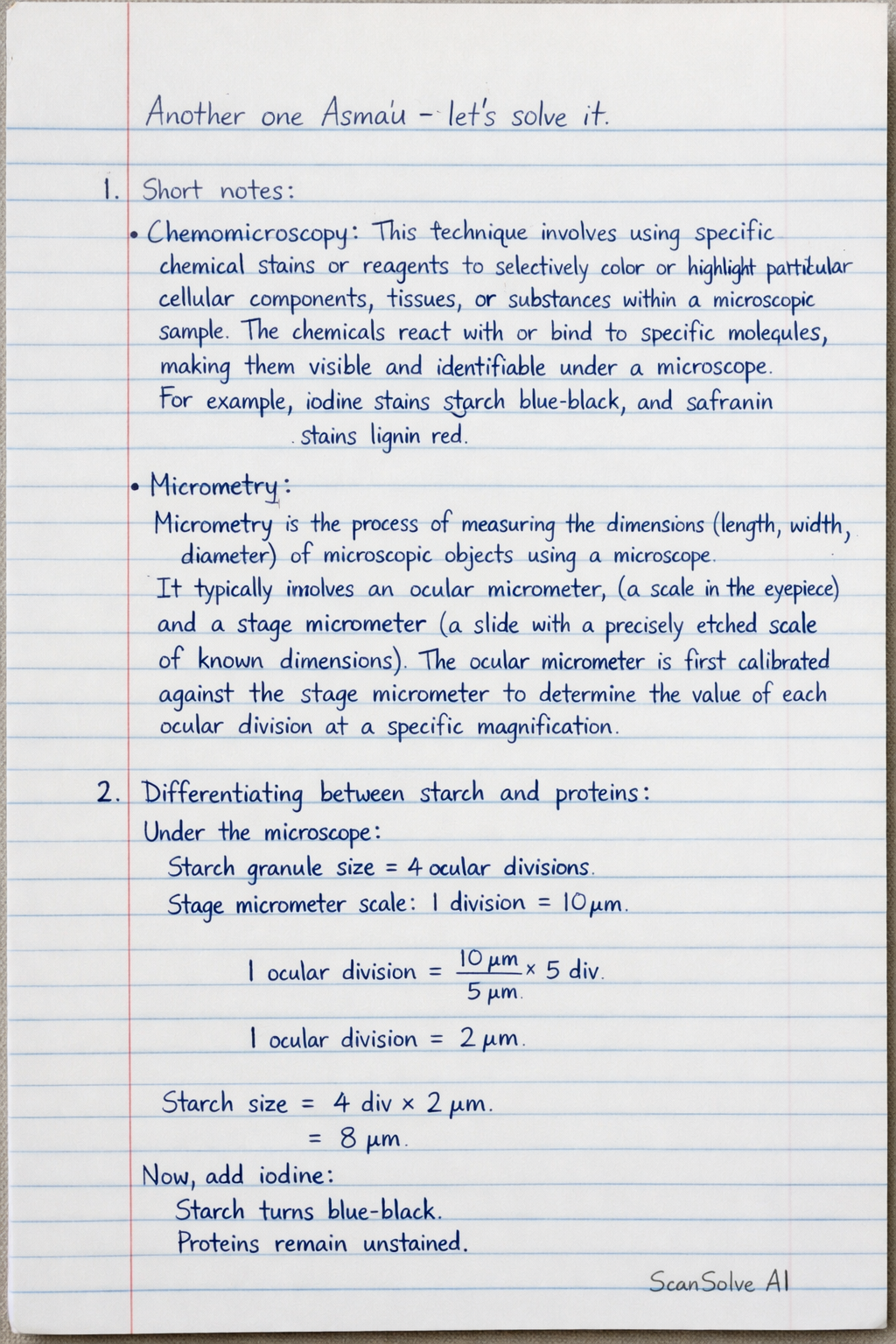
Another one Asma'u — let's solve it. 1. Short notes: Chemomicroscopy*: This technique involves using specific chemical stains or reagents to selectively color or highlight particular cellular components, tissues, or substances within a microscopic sample. The chemicals react with or bind to specific molecules, making them visible and identifiable under a microscope. For example, iodine stains starch blue-black, and safranin stains lignin red. Micrometry: Micrometry is the process of measuring the dimensions (length, width, diameter) of microscopic objects using a microscope. It typically involves an ocular micrometer (a scale in the eyepiece) and a stage micrometer* (a slide with a precisely etched scale of known dimensions). The ocular micrometer is first calibrated against the stage micrometer to determine the value of each ocular division at a specific magnification. 2. Differentiating between starch and proteins: Physical Means: Starch*: Typically appears as a white, amorphous powder. It is insoluble in cold water but forms a viscous paste or gel when heated in water. Proteins*: Can exist as white powders, crystals, or amorphous solids. Their solubility in water varies greatly; some are highly soluble, while others are insoluble. Chemical Means: Starch: The iodine test* is used. When a few drops of iodine solution are added to starch, it produces a characteristic blue-black color due to the formation of an inclusion complex between iodine and the coiled amylose molecules in starch. Proteins: The Biuret test* is commonly used. When a protein solution is mixed with a strong base (like NaOH) and then a dilute copper(II) sulfate solution, a violet or purple color develops. This color indicates the presence of peptide bonds (at least two) in the protein. 3. Short notes on stomata types: Paracytic stomata*: In this type of stomata, the guard cells are accompanied by one or more subsidiary cells that are arranged parallel to the long axis of the guard cells and the stomatal pore. This arrangement is characteristic of plants like coffee (Coffea) and members of the Rubiaceae family. Anisocytic stomata*: Also known as cruciferous stomata, this type is characterized by the guard cells being surrounded by three subsidiary cells of unequal size, with one being distinctly smaller than the other two. This arrangement is common in plants belonging to the Brassicaceae (mustard) family. 4. Calibration factor calculation: To calculate the calibration factor, we compare the divisions of an ocular micrometer (OM) with a stage micrometer (SM) at a specific magnification. Step 1: Understand the stage micrometer. A common stage micrometer has a scale where 1 \, mm is divided into 100 parts. Therefore, each division on the stage micrometer represents: 1 \, mm100 = 0.01 \, mm = 10 \, Step 2: Assume alignment between ocular and stage micrometers. Let's assume that 10 divisions on the ocular micrometer align perfectly with 4 divisions on the stage micrometer. Step 3: Calculate the total length represented by the aligned stage micrometer divisions. 4 \, SM divisions = 4 × 10 \, = 40 \, Step 4: Determine the value of one ocular micrometer division (the calibration factor). Since 10 ocular divisions correspond to 40 \, : 1 \, OM division = 40 \, 10 \, OM divisions = 4 \, /OM division The calibration factor is 4 \, . What's next? Send 'em! 📸