
We read your photo as:
Differentiate basic and acid dyes. 52. List three histological methods of studying living tissues and define any.
ScanSolve AI Answer
Here are the answers to your questions:
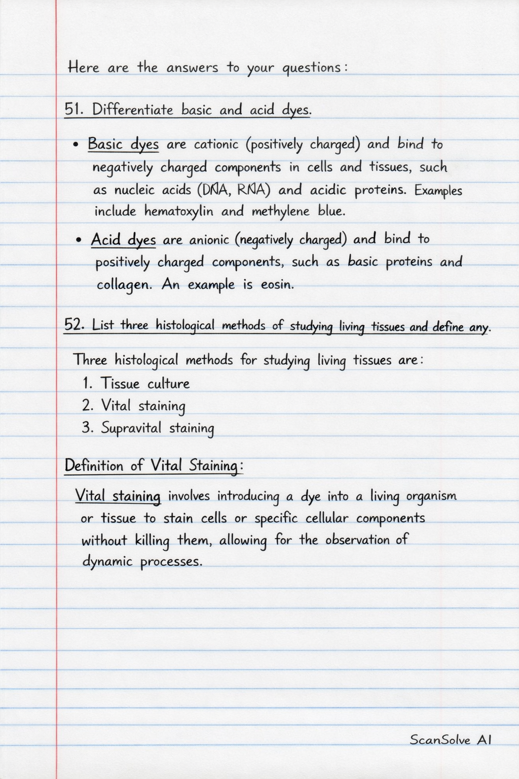
51. Differentiate basic and acid dyes.
- Basic dyes are cationic (positively charged) and bind to negatively charged components in cells and tissues, such as nucleic acids (DNA, RNA) and acidic proteins. Examples include hematoxylin and methylene blue.
- Acid dyes are anionic (negatively charged) and bind to positively charged components, such as basic proteins and collagen. An example is eosin.
52. List three histological methods of studying living tissues and define any.
Three histological methods for studying living tissues are:
- Tissue culture
- Vital staining
- Supravital staining
Definition of Vital Staining:
Vital staining involves introducing a dye into a living organism or tissue to stain cells or specific cellular components without killing them, allowing for the observation of dynamic processes.
53. Outline the advantages of using killed chemically preserved tissue in histology.
The advantages of using killed chemically preserved tissue in histology include:
- Prevention of autolysis and putrefaction: Chemical fixatives stop enzymatic degradation and bacterial decomposition.
- Preservation of tissue structure: Fixation stabilizes cellular and tissue components, maintaining their morphology.
- Long-term storage: Preserved tissues can be stored for extended periods without degradation.
- Enhanced tissue rigidity: Fixation makes tissues firmer, facilitating easier sectioning.
- Inactivation of pathogens: Fixatives kill bacteria and viruses, making tissues safer to handle.
54. Define histochemistry and cytochemistry.
- Histochemistry is the branch of histology that uses specific chemical reactions to identify and localize various chemical substances within tissues.
- Cytochemistry is a specialized area of histochemistry that focuses on identifying and localizing chemical substances within individual cells.
55. State the four types of somatic tissues, location in organs and functions.
The four primary types of somatic tissues are:
-
1. Epithelial Tissue
- Location: Covers body surfaces, lines hollow organs, body cavities, and ducts; forms glands.
- Functions: Protection, secretion, absorption, filtration, and sensory reception.
-
2. Connective Tissue
- Location: Widely distributed throughout the body; found in bones, cartilage, fat, blood, ligaments, and tendons.
- Functions: Supports, protects, binds other tissues together, stores energy, and transports substances.
-
3. Muscle Tissue
- Location: Skeletal muscles (attached to bones), smooth muscles (walls of internal organs), and cardiac muscle (heart).
- Functions: Movement, maintenance of posture, and heat production.
-
4. Nervous Tissue
- Location: Brain, spinal cord, and nerves.
- Functions: Transmits electrical signals, coordinates, and controls body activities.
Still stuck on this one?
Ask a follow-up, and the answer starts from this question.
More Biology Questions
Handwritten Step-by-Step Solution
Get this as a handwritten image on WhatsApp — like a tutor wrote it for you.
Try on WhatsApp