
We read your photo as:
Describe and illustrate the effect of distilled water and 1M NaCl on plant cells, labeling all key organelles and cell structures.
ScanSolve AI Answer
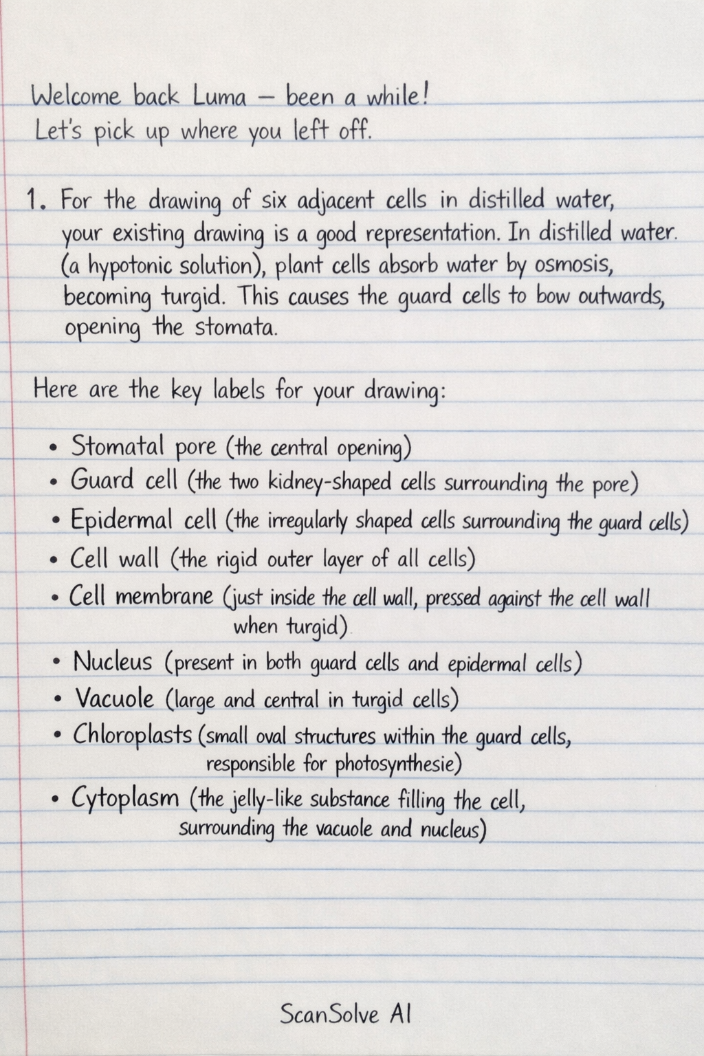
1. For the drawing of six adjacent cells in distilled water, your existing drawing is a good representation. In distilled water (a hypotonic solution), plant cells absorb water by osmosis, becoming turgid. This causes the guard cells to bow outwards, opening the stomata.
Here are the key labels for your drawing:
- Stomatal pore (the central opening)
- Guard cell (the two kidney-shaped cells surrounding the pore)
- Epidermal cell (the irregularly shaped cells surrounding the guard cells)
- Cell wall (the rigid outer layer of all cells)
- Cell membrane (just inside the cell wall, pressed against the cell wall when turgid)
- Nucleus (present in both guard cells and epidermal cells)
- Vacuole (large and central in turgid cells)
- Chloroplasts (small oval structures within the guard cells, responsible for photosynthesis)
- Cytoplasm (the jelly-like substance filling the cell, surrounding the vacuole and nucleus)
- Thick inner wall of guard cell (the wall facing the stomatal pore)
- Thin outer wall of guard cell (the wall facing away from the stomatal pore)
m. For the labelled drawing of six adjacent cells in 1M NaCl, you need to show the effects of a hypertonic solution. In 1M NaCl, water will move out of the cells by osmosis, causing them to lose turgor and undergo plasmolysis.
Here is a description of what your drawing should depict:
- The stomatal pore should be closed or nearly closed, as the guard cells will have lost turgor and become flaccid.
- The guard cells should appear flaccid and less bowed, possibly straighter.
- The epidermal cells should show clear plasmolysis. This means the cell membrane and the entire protoplast (cytoplasm, nucleus, and vacuole) will have shrunk and pulled away from the rigid cell wall. The cell wall itself will retain its original shape.
- The vacuoles within all cells will appear smaller and shrunken.
- The nucleus and chloroplasts (in guard cells) will be visible within the shrunken protoplast.
Here are the key labels for this drawing:
- Closed stomatal pore
- Flaccid guard cell
- Plasmolysed epidermal cell
- Cell wall (retaining original shape)
- Cell membrane (pulled away from the cell wall)
- Shrunken protoplast (the shrunken contents of the cell, including cytoplasm, nucleus, and vacuole)
- Nucleus
- Shrunken vacuole
- Chloroplasts (in guard cells, within the shrunken protoplast)
Still stuck on this one?
Ask a follow-up, and the answer starts from this question.
More Biology Questions
Handwritten Step-by-Step Solution
Get this as a handwritten image on WhatsApp — like a tutor wrote it for you.
Try on WhatsApp