

This biology question covers important biological concepts and processes. The step-by-step explanation below helps you understand the underlying mechanisms and reasoning.
ScanSolve AI Answer
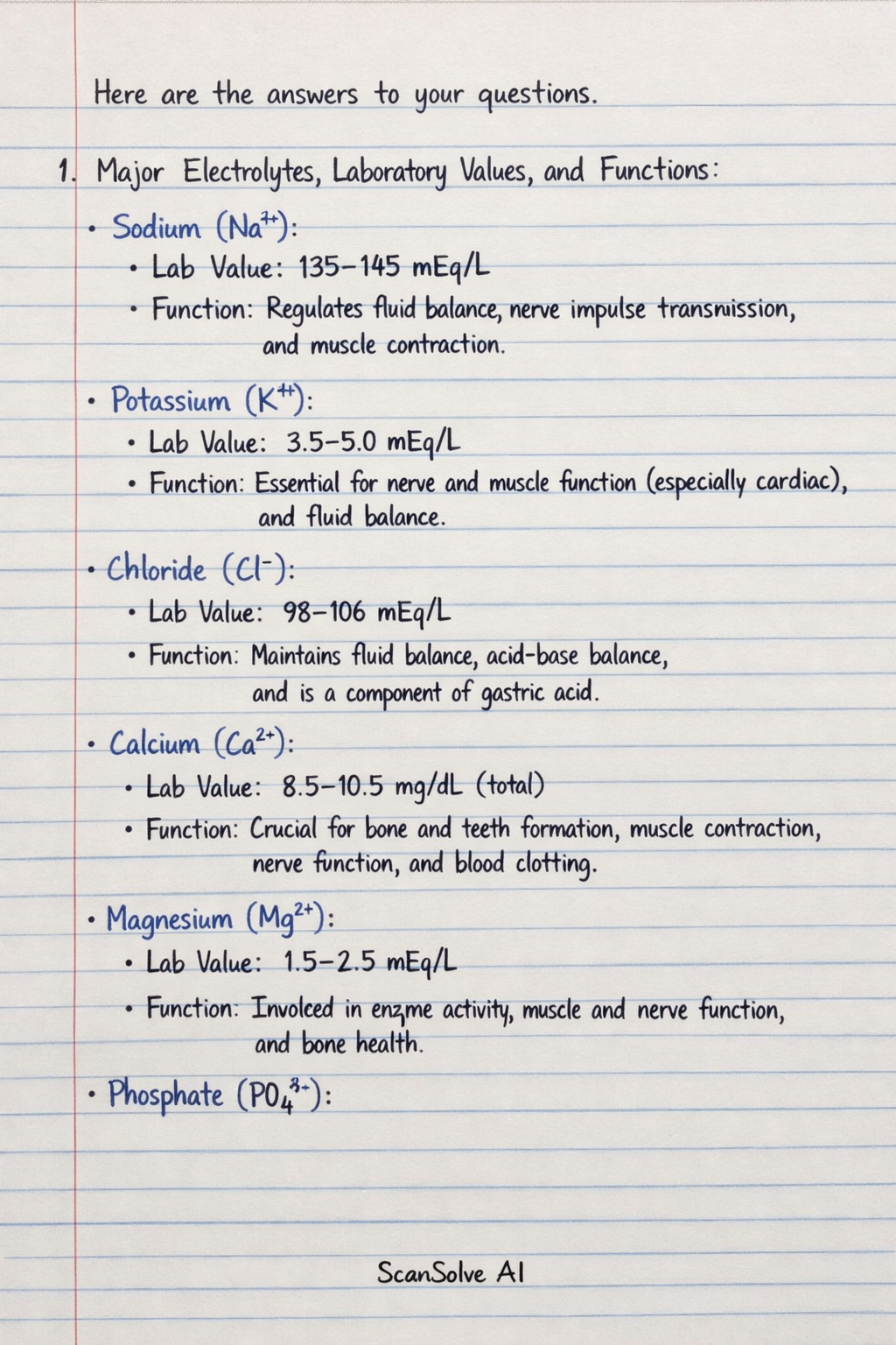
Here are the answers to your questions. 1. Major Electrolytes, Laboratory Values, and Functions: Sodium (Na^+) Lab Value: 135-145 mEq/L Function: Regulates fluid balance, nerve impulse transmission, and muscle contraction. Potassium (K^+) Lab Value: 3.5-5.0 mEq/L Function: Essential for nerve and muscle function (especially cardiac), and fluid balance. Chloride (Cl^-) Lab Value: 98-106 mEq/L Function: Maintains fluid balance, acid-base balance, and is a component of gastric acid. Calcium (Ca^2+) Lab Value: 8.5-10.5 mg/dL (total) Function: Crucial for bone and teeth formation, muscle contraction, nerve function, and blood clotting. Magnesium (Mg^2+) Lab Value: 1.5-2.5 mEq/L Function: Involved in enzyme activity, muscle and nerve function, and bone health. Phosphate (PO_4^3-) Lab Value: 2.5-4.5 mg/dL Function: Important for bone and teeth formation, energy storage (ATP), and acid-base balance. Bicarbonate (HCO_3^-) Lab Value: 22-28 mEq/L Function: A primary buffer in the body's acid-base balance system. 2. Three Negative Feedback Mechanisms and Involved Organs: Regulation of Blood Glucose Mechanism: When blood glucose levels rise, the pancreas releases insulin, which promotes glucose uptake by liver, muscle, and adipose cells, lowering blood glucose. When levels fall, the pancreas releases glucagon, which signals the liver* to release stored glucose. Organs: Pancreas, liver, muscle cells, adipose tissue*. Regulation of Body Temperature Mechanism: The hypothalamus detects changes in body temperature. If temperature rises, it triggers sweat glands to produce sweat and blood vessels in the skin to dilate. If temperature falls, it triggers muscles to shiver and blood vessels* to constrict. Organs: Hypothalamus (brain), skin, sweat glands, blood vessels, muscles*. Regulation of Thyroid Hormones Mechanism: High levels of thyroid hormones (T3 and T4) inhibit the release of Thyroid-Stimulating Hormone (TSH) from the anterior pituitary gland and Thyrotropin-Releasing Hormone (TRH) from the hypothalamus. Low levels stimulate their release, leading to increased thyroid hormone production by the thyroid gland*. Organs: Hypothalamus, anterior pituitary gland, thyroid gland*. 3. Cell Division, Organelles, and Differentiation between Mitosis and Meiosis: Process of Cell Division (General Explanation) Cell division is the process by which a parent cell divides into two or more daughter cells. It typically involves two main phases: interphase, where the cell grows and duplicates its DNA, and the M phase* (mitotic or meiotic phase), where the cell divides its nucleus (karyokinesis) and then its cytoplasm (cytokinesis). Note: As an AI, I cannot provide a diagram. A typical diagram would illustrate the stages of mitosis or meiosis with chromosomes and cellular structures.* Cell Organelles Involved in Cell Division Nucleus*: Contains the cell's genetic material (DNA) which is replicated and segregated. Centrosomes/Centrioles*: Organize the mitotic or meiotic spindle fibers, which are crucial for chromosome movement. Ribosomes*: Synthesize proteins necessary for cell growth and division. Mitochondria*: Provide the ATP (energy) required for the energetic processes of cell division. Endoplasmic Reticulum and Golgi Apparatus*: Involved in synthesizing and processing membranes and proteins needed for new daughter cells. Differentiation between Mitosis and Meiosis Mitosis: Occurs in somatic cells*. Involves one* round of cell division. Results in two diploid (2n)* daughter cells. Daughter cells are genetically identical* to the parent cell. Function: Growth, repair, and asexual reproduction. Meiosis: Occurs in germ cells* (for sexual reproduction). Involves two* rounds of cell division (Meiosis I and Meiosis II). Results in four haploid (n)* daughter cells. Daughter cells are genetically diverse* from the parent cell due to crossing over and independent assortment. Function: Production of gametes (sperm and egg cells). 4. Process of Meiosis: Note: As an AI, I cannot provide a diagram. A typical diagram would illustrate the two divisions, Meiosis I and Meiosis II, showing chromosome pairing, crossing over, and segregation.* Meiosis I (Reductional Division): Prophase I: Chromosomes condense, homologous chromosomes pair up to form bivalents, and crossing over* occurs (exchange of genetic material). The nuclear envelope breaks down, and the spindle fibers begin to form. Metaphase I: Homologous chromosome pairs align along the metaphase plate. Anaphase I: Homologous chromosomes separate and move to opposite poles of the cell. Sister chromatids remain attached. Telophase I & Cytokinesis: Chromosomes decondense, nuclear envelopes may reform, and the cytoplasm divides, resulting in two haploid cells, each with duplicated chromosomes. Meiosis II (Equational Division): Prophase II: Chromosomes condense again, and the nuclear envelope (if reformed) breaks down. Spindle fibers form. Metaphase II: Sister chromatids align along the metaphase plate in each of the two cells. Anaphase II: Sister chromatids separate and move to opposite poles, becoming individual chromosomes. Telophase II & Cytokinesis: Chromosomes decondense, nuclear envelopes reform around the four sets of chromosomes, and the cytoplasm divides. This results in four haploid daughter cells*, each with unduplicated chromosomes. 5. Structures Cut During a Cesarean Section (from skin to baby): 1. Skin 2. Subcutaneous tissue (fat) 3. Anterior rectus sheath (a layer of fascia) 4. Peritoneum (parietal and visceral layers, often entered as one) 5. Uterine wall (myometrium) 6. Amniotic sac (membranes) 7. Baby