
We read your photo as:
Describe the staining reactions of secretory cells and the different types and modes of glandular secretion.
ScanSolve AI Answer
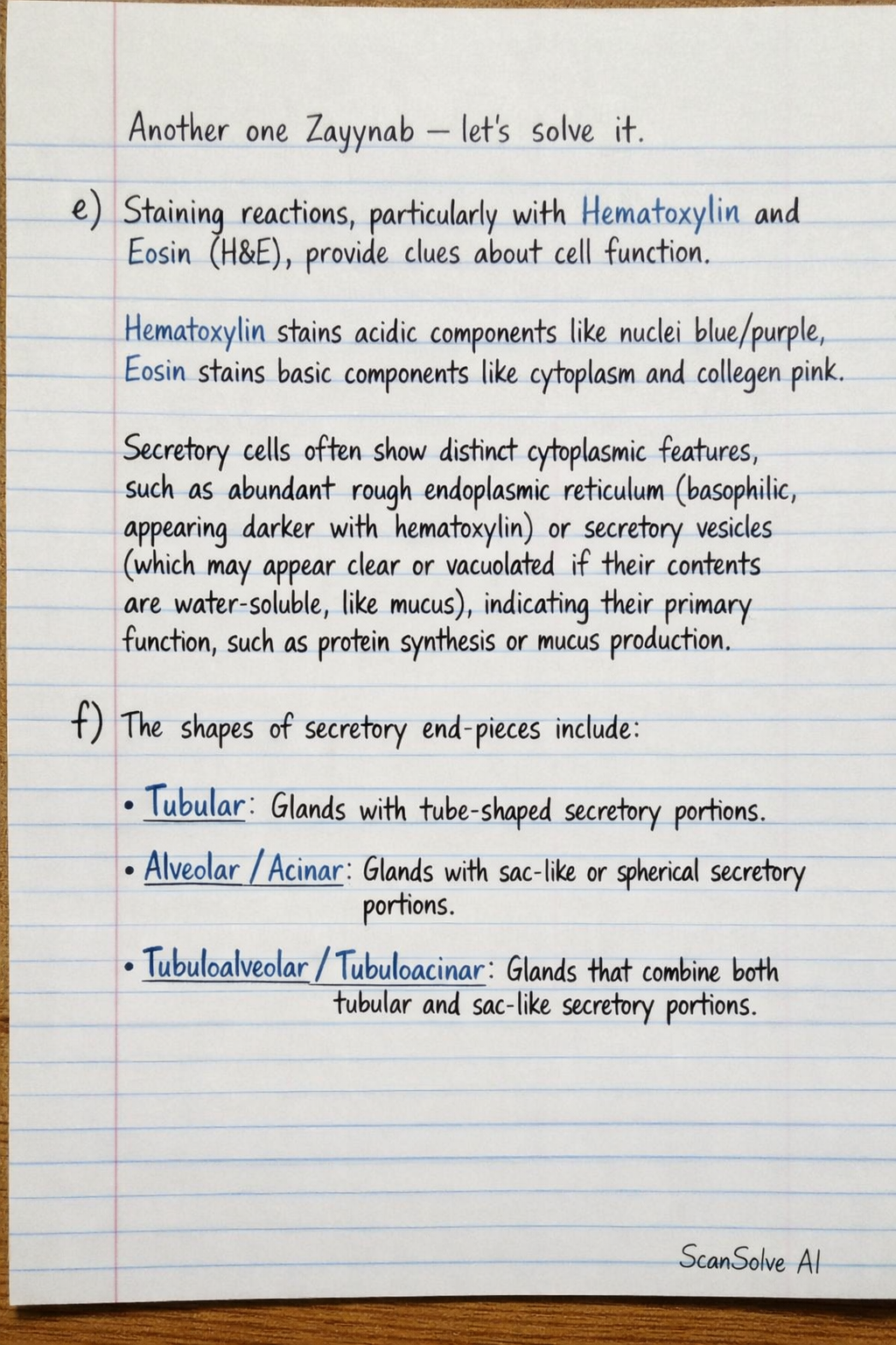
e) Staining reactions, particularly with Hematoxylin and Eosin (H&E), provide clues about cell function. Hematoxylin stains acidic components like nuclei blue/purple, while Eosin stains basic components like cytoplasm and collagen pink. Secretory cells often show distinct cytoplasmic features, such as abundant rough endoplasmic reticulum (basophilic, appearing darker with hematoxylin) or secretory vesicles (which may appear clear or vacuolated if their contents are water-soluble, like mucus), indicating their primary function, such as protein synthesis or mucus production.
f)
The shapes of secretory end-pieces include:
- Tubular: Glands with tube-shaped secretory portions.
- Alveolar/Acinar: Glands with sac-like or spherical secretory portions.
- Tubuloalveolar/Tubuloacinar: Glands that combine both tubular and sac-like secretory portions.
The modes of secretion are:
- Merocrine secretion: Secretory products are released from the cell via exocytosis without loss of cellular material.
- Apocrine secretion: Secretory products accumulate at the apical surface, and a portion of the cytoplasm, along with the product, is pinched off.
- Holocrine secretion: The entire cell fills with secretory product, then disintegrates, releasing the product and cell debris.
I. UNICELLULAR GLANDS (Goblet Cells)
Goblet cells are unicellular glands found scattered within the simple columnar epithelium of the small intestines and respiratory tract. They are specialized for producing and secreting mucus. In routine H&E staining, the mucinogen granules within goblet cells are water-soluble and often wash out during tissue processing, causing the cells to appear pale-staining or clear and distended, resembling a goblet. The nucleus is typically compressed and located at the base of the cell. A drawing would illustrate these clear, goblet-shaped cells interspersed among the taller, darker-staining columnar epithelial cells, with their basal nuclei.
Still stuck on this one?
Ask a follow-up, and the answer starts from this question.
More Biology Questions
Handwritten Step-by-Step Solution
Get this as a handwritten image on WhatsApp — like a tutor wrote it for you.
Try on WhatsApp