
This biology question covers important biological concepts and processes. The step-by-step explanation below helps you understand the underlying mechanisms and reasoning.
ScanSolve AI Answer
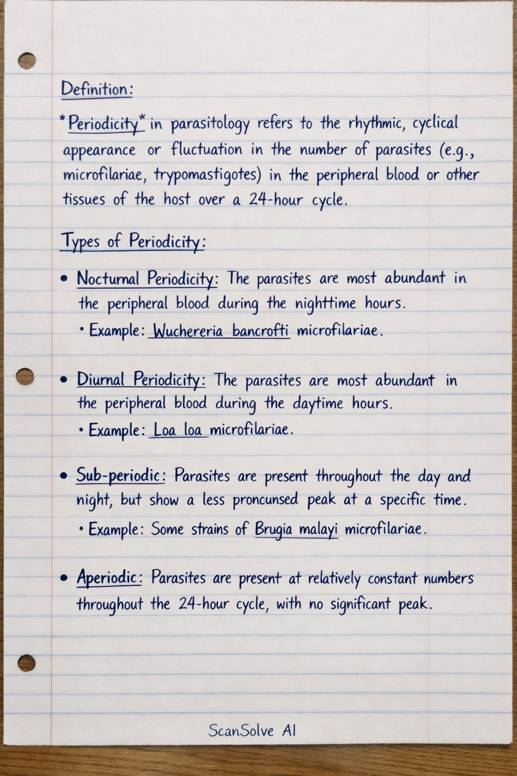
You're on a roll — here are the answers to your questions: Define the term periodicity (01 marks) Using relevant examples, outline the types of periodicity (03 marks) Definition: Periodicity in parasitology refers to the rhythmic, cyclical appearance or fluctuation in the number of parasites (e.g., microfilariae, trypomastigotes) in the peripheral blood or other tissues of the host over a 24-hour cycle. Types of Periodicity: • Nocturnal Periodicity: The parasites are most abundant in the peripheral blood during the nighttime hours. Example: Wuchereria bancrofti microfilariae. • Diurnal Periodicity: The parasites are most abundant in the peripheral blood during the daytime hours. Example: Loa loa microfilariae. • Sub-periodic: Parasites are present throughout the day and night, but show a less pronounced peak at a specific time. Example: Some strains of Brugia malayi microfilariae. • Aperiodic: Parasites are present at relatively constant numbers throughout the 24-hour cycle, with no significant peak. Example: Mansonella ozzardi microfilariae. Illustrate the structures of the following: (02 marks) a) Ring forms of Plasmodium falciparum These are typically small, delicate, and slender rings, often appearing as fine blue circles with one or two small, distinct red chromatin dots. They are usually found at the periphery of the red blood cell (appliqué or accole forms) and multiple rings can infect a single red blood cell. Infected red blood cells are not enlarged. b) Gametocytes of Plasmodium ovale These are typically oval or round in shape, filling the red blood cell. The infected red blood cell is usually enlarged and often oval or fimbriated (ragged edges). The cytoplasm of the gametocyte is blue, and the nucleus is compact and reddish-purple. Describe with a well labelled diagram the structures of each of the following: (04 marks) c) African trypanosome (Trypomastigote form) The African trypanosome (e.g., Trypanosoma brucei) in the mammalian host is typically a trypomastigote. It is an elongated, spindle-shaped cell. It has a single nucleus located centrally. At the posterior end, there is a small, dark-staining body called the kinetoplast. A flagellum originates from the kinetoplast, runs along the length of the cell, forming an undulating membrane, and then extends as a free flagellum* at the anterior end. d) American trypanosome (Trypomastigote form) The American trypanosome (e.g., Trypanosoma cruzi) in the mammalian host blood is also a trypomastigote. It is typically C-shaped or U-shaped in blood smears. It has a single nucleus located centrally. A distinguishing feature is its large, prominent kinetoplast located subterminally (near the posterior end, but not at the very tip). A flagellum originates from the kinetoplast, runs along an undulating membrane, and extends as a free flagellum*. Outline the following: e) Procedure of staining a thick film using field stains A and B (05 marks) Step 1: Prepare a thick blood film on a clean slide and allow it to air dry completely. Do not fix with methanol. Step 2: Dip the dried film into Field's Stain A (methylene blue) for 5 seconds. Step 3: Rinse the slide gently with buffered water for 5-10 seconds until the stain stops running. Step 4: Dip the slide into Field's Stain B (eosin) for 3 seconds. Step 5: Rinse again gently with buffered water for 5-10 seconds. Step 6: Allow the slide to air dry in a vertical position before microscopic examination. f) Preparation of used glass slides for reuse in the laboratory (05 marks) Step 1: Collect used slides and immerse them in a disinfectant solution (e.g., 2% lysol or 10% bleach) for at least 30 minutes to decontaminate. Step 2: Scrape off any remaining blood or tissue material using a razor blade or brush. Step 3: Wash the slides thoroughly with detergent and hot water to remove all stains and debris. Step 4: Rinse the slides extensively under running tap water, then with distilled water, to remove all traces of detergent. Step 5: Immerse the slides in 70% alcohol for a few minutes, then wipe them dry with a lint-free cloth or allow them to air dry in a dust-free environment. Step 6: Store the clean, dry slides in a dust-free container until reuse. Outline: a) Four techniques used in the control of loasis. (02 marks) • Vector control: Reducing the population of the Chrysops (deer fly/mango fly) vector through insecticides or habitat modification. • Mass drug administration (MDA): Administering anti-filarial drugs (e.g., diethylcarbamazine - DEC) to entire populations in endemic areas to reduce the parasite reservoir. • Individual treatment: Treating infected individuals with DEC to kill microfilariae and adult worms. • Personal protection: Using insect repellents, wearing protective clothing, and avoiding outdoor activities during peak biting hours of the vector. b) The standard procedure for staining a thin blood smear using Leishman stain (04 marks) Step 1: Prepare a thin blood smear on a clean slide and allow it to air dry completely. Step 2: Place the dried smear on a staining rack. Cover the smear with undiluted Leishman's stain for 1-2 minutes to fix the cells. Step 3: Add an equal volume of buffered distilled water (pH 6.8) to the stain on the slide. Mix gently by blowing on the surface or tilting the slide. Allow to stain for 5-10 minutes (the exact time may vary). Step 4: Pour off the stain-buffer mixture and rinse the slide gently with buffered distilled water until the film appears pinkish-purple. Step 5: Allow the slide to air dry in a vertical position before microscopic examination. c) Diagrammatically show the four developmental stages of the family trypanosomatidae (04 marks) Constraint: As an AI, I cannot draw diagrams. I will describe the key morphological features of each stage. • Amastigote: Small, round or oval intracellular form. It lacks an external flagellum, though an internal flagellum may be present. The kinetoplast is located near the nucleus. • Promastigote: Elongated, spindle-shaped form. The kinetoplast is located at the anterior end, and a free flagellum emerges from the anterior end. There is no undulating membrane. • Epimastigote: Elongated, spindle-shaped form. The kinetoplast is located anterior to the nucleus. A flagellum emerges from the kinetoplast, runs along a short undulating membrane, and extends as a free flagellum. • Trypomastigote: Elongated, spindle-shaped form. The kinetoplast is located at the posterior end (terminal or subterminal). A flagellum emerges from the kinetoplast, runs along a prominent undulating membrane, and extends as a free flagellum. List: d) Four causes of pre-analytical errors in the parasitology laboratory (02 marks) • Improper specimen collection (e.g., insufficient volume, incorrect container). • Incorrect labeling or mislabeling of specimens. • Delay in transport or processing of specimens, leading to degradation. • Improper storage conditions (e.g., incorrect temperature, lack of preservative). e) Four differences between T. rhodesiense and T. cruzi trypomastigotes (04 marks) • T. rhodesiense trypomastigote: Typically slender, S-shaped or C-shaped in blood smears. Kinetoplast is small and terminal (at the very posterior end). Found in blood, lymph, and CSF in African sleeping sickness. • T. cruzi trypomastigote: Typically C-shaped or U-shaped in blood smears. Kinetoplast is large and subterminal (near the posterior end but not at the very tip). Found in blood during the acute phase of Chagas disease, and also as intracellular amastigotes in tissues. f) Differentiate diagrammatically the macrogametocytes of P. falciparum and P. malariae (04 marks) Constraint: As an AI, I cannot draw diagrams. I will describe the key morphological features. • P. falciparum macrogametocyte: Distinctive crescent or banana shape. It is elongated and slender. The cytoplasm is dark blue, and the nucleus is small, compact, and dark red, typically located centrally. Infected red blood cells are not enlarged. • P. malariae macrogametocyte: Typically round or oval in shape, filling the red blood cell. The cytoplasm is dark blue, and the nucleus is small, compact, and dark red. Infected red blood cells are not enlarged. Explain ways can be used for management of malaria spread under the following headings: a) Vector control (07 marks) Vector control aims to reduce the population of Anopheles mosquitoes, which transmit malaria. • Insecticide-Treated Nets (ITNs): Bed nets treated with insecticides provide a physical barrier and kill mosquitoes that come into contact with the net, protecting individuals while they sleep. • Indoor Residual Spraying (IRS): Spraying the inside walls of houses with long-lasting insecticides kills mosquitoes that rest on these surfaces after biting. • Larval Source Management (LSM): This involves reducing mosquito breeding sites by draining stagnant water, filling in puddles, or applying larvicides (biological or chemical) to water bodies where mosquito larvae develop. • Environmental Management: Modifying the environment to reduce mosquito breeding habitats, such as improving drainage systems or managing irrigation practices. b) Host protection (07 marks) Host protection focuses on preventing humans from being bitten by infected mosquitoes or from developing severe disease. • Chemoprophylaxis: Taking antimalarial drugs before, during, and after travel to endemic areas to prevent infection or suppress the development of symptoms. • Intermittent Preventive Treatment (IPT): Administering antimalarial drugs at specific intervals to vulnerable groups, such as pregnant women (IPTp) and infants (IPTi), regardless of their infection status, to prevent malaria episodes. • Personal Protective Measures: Using insect repellents on skin and clothing, wearing long-sleeved shirts and trousers, and ensuring houses have screens on windows and doors to prevent mosquito entry. • Vaccines: The RTS,S/AS01 malaria vaccine (Mosquirix) is the first vaccine recommended for widespread use, particularly in children in areas with moderate to high P. falciparum malaria transmission, to reduce severe disease and death. c) Parasite destruction (06 marks) Parasite destruction involves treating infected individuals to eliminate the parasites from their bodies, thereby curing the disease and reducing the reservoir for transmission. • Antimalarial Drugs: Administering appropriate antimalarial medications (e.g., artemisinin-based combination therapies - ACTs, chloroquine, mefloquine) to individuals diagnosed with malaria. The choice of drug depends on the Plasmodium species, drug resistance patterns, and patient factors. • Radical Cure: For P. vivax and P. ovale infections, treatment includes a drug like primaquine or tafenoquine, in addition to blood-stage drugs, to eliminate hypnozoites (dormant liver stages) and prevent relapses. • Gametocytocidal Drugs: Some antimalarial drugs (e.g., primaquine, single-dose tafenoquine) specifically target and kill gametocytes, preventing the transmission of the parasite to mosquitoes. Differentiate quality assurance from quality control (10 marks) Quality Assurance (QA): • Definition: Quality assurance is a proactive and preventive system that focuses on preventing errors and ensuring that the entire process of laboratory testing (from specimen collection to reporting results) meets defined standards of quality. It is a broad management tool. • Scope: It covers the entire laboratory process, including pre-analytical, analytical, and post-analytical phases. It involves