
We read your photo as:
A) Describe the procedure for formol ether concentration technique/method
ScanSolve AI Answer
Step-by-step solution
7 stepsHere's the procedure for the formol-ether concentration technique and how to interpret and report the results:
Part a· 7 steps
Describe the procedure for formol ether concentration technique/method
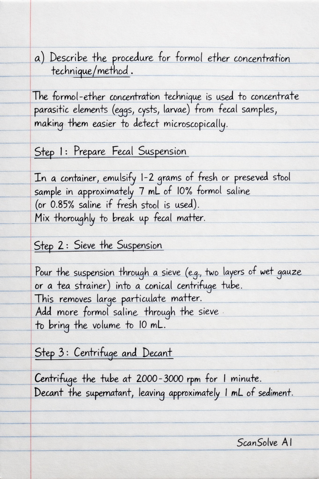
The formol-ether concentration technique is used to concentrate parasitic elements (eggs, cysts, larvae) from fecal samples, making them easier to detect microscopically.
Prepare Fecal Suspension
In a container, emulsify 1-2 grams of fresh or preserved stool sample in approximately 7 mL of 10% formol saline (or 0.85% saline if fresh stool is used). Mix thoroughly to break up fecal matter.
Sieve the Suspension
Pour the suspension through a sieve (e.g., two layers of wet gauze or a tea strainer) into a conical centrifuge tube. This removes large particulate matter. Add more formol saline through the sieve to bring the volume to 10 mL.
Centrifuge and Decant
Centrifuge the tube at 2000-3000 rpm for 1 minute. Decant the supernatant, leaving approximately 1 mL of sediment.
Wash Sediment (Optional but Recommended)
Resuspend the sediment in 10 mL of 10% formol saline. Centrifuge again at 2000-3000 rpm for 1 minute and decant the supernatant. Repeat this wash step if the supernatant is still very cloudy.
Add Formalin and Ether
Add 7 mL of 10% formol saline to the sediment, mix well. Then, add 3 mL of diethyl ether (or ethyl acetate) to the tube. Cap the tube and shake vigorously for 30 seconds to 1 minute.
Centrifuge for Separation
Centrifuge the mixture at 2000-3000 rpm for 1 minute. Four layers will form:
- Top layer: Ether
- Second layer: Fecal debris plug
- Third layer: Formalin
- Bottom layer: Sediment containing parasites
Decant and Prepare Mount
Carefully loosen the fecal debris plug with an applicator stick. Quickly invert the tube to decant the top three layers, leaving only the concentrated sediment at the bottom. Use a pipette to transfer a small drop of the sediment onto a clean microscope slide. Add a drop of Lugol's iodine or saline, cover with a coverslip, and examine microscopically.
Part b
Write the Interpretation and Reporting Results
Interpretation and reporting involve identifying any parasitic forms found in the concentrated sediment.
- Microscopic Examination: Systematically scan the entire wet mount under low power (100x) to locate larger elements like helminth eggs and larvae. Then, switch to high power (400x) to identify protozoan cysts and trophozoites (if present and viable) and confirm the morphology of helminth eggs.
- Identification: Identify parasites based on their characteristic morphology, size, and internal structures. Use reference images or keys for confirmation.
- Reporting:
- Report the presence or absence of specific parasites.
- If parasites are found, report the species identified (e.g., Ascaris lumbricoides eggs, Giardia lamblia cysts, Entamoeba histolytica cysts).
- For quantitative reporting, an estimate of the number of parasites (e.g., "few," "moderate," "many") can be included, though this method is primarily qualitative.
- A typical report might state: "Concentrated stool examination: Entamoeba coli cysts present (few)" or "Concentrated stool examination: No parasites seen."
Still stuck on this one?
Ask a follow-up, and the answer starts from this question.
More Chemistry Questions
Handwritten Step-by-Step Solution
Get this as a handwritten image on WhatsApp — like a tutor wrote it for you.
Try on WhatsApp