
We read your photo as:
Define and explain Wuchereria bancrofti, Mode of transmission and pathogenesis, Pathogenicity and clinical features, Diagnosis, Treatment, Life cycle.
Biology
ScanSolve AI Answer
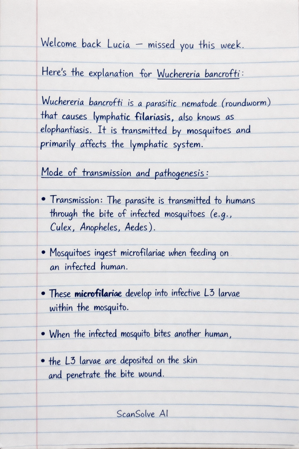
Here's the explanation for Wuchereria bancrofti:
Wuchereria bancrofti is a parasitic nematode (roundworm) that causes lymphatic filariasis, also known as elephantiasis. It is transmitted by mosquitoes and primarily affects the lymphatic system.
-
Mode of transmission and pathogenesis:
- Transmission: The parasite is transmitted to humans through the bite of infected mosquitoes (e.g., Culex, Anopheles, Aedes). Mosquitoes ingest microfilariae when feeding on an infected human. These microfilariae develop into infective L3 larvae within the mosquito. When the infected mosquito bites another human, the L3 larvae are deposited on the skin and penetrate the bite wound.
- Pathogenesis: Once inside the human host, the L3 larvae migrate to the lymphatic vessels and lymph nodes, where they mature into adult worms. These adult worms can live for many years, producing millions of microfilariae that circulate in the blood, primarily at night. The presence of adult worms in the lymphatic system causes inflammation, obstruction, and damage, leading to the characteristic symptoms of lymphatic filariasis.
-
Pathogenicity and clinical features:
- Pathogenicity: The adult worms cause lymphatic dysfunction, leading to lymphedema (swelling due to fluid accumulation). This chronic inflammation and obstruction can lead to fibrosis and thickening of the lymphatic vessels.
- Clinical features:
- Acute manifestations: Fever, lymphadenitis (inflammation of lymph nodes), and funiculitis (inflammation of the spermatic cord).
- Chronic manifestations: Lymphedema (swelling of limbs, breasts, or genitalia), hydrocele (fluid accumulation in the scrotum), and elephantiasis (severe thickening and hardening of the skin and underlying tissues, typically in the legs or scrotum).
-
Diagnosis:
- The primary diagnostic method is the microscopic detection of microfilariae in blood smears. Blood samples are usually collected at night (between 10 PM and 2 AM) because microfilariae exhibit nocturnal periodicity.
- Antigen detection tests (immunochromatographic card tests) are also available and can detect circulating filarial antigens, which are present even when microfilariae are not detectable.
- Ultrasound can be used to visualize adult worms in the lymphatic vessels, often referred to as the "filarial dance sign."
-
Treatment:
- The main drugs used are Diethylcarbamazine (DEC), Albendazole, and Ivermectin. These drugs kill microfilariae and, to some extent, adult worms.
- Mass drug administration (MDA) programs are often implemented in endemic areas to reduce the parasite load in the community.
- Surgical intervention may be necessary for chronic conditions like hydrocele or to manage severe elephantiasis.
-
Life Cycle:
- Mosquito takes a blood meal: Injects L3 larvae onto the human skin.
- Larvae enter human: L3 larvae penetrate the skin and migrate to lymphatic vessels.
- Maturation: Larvae develop into adult worms in the lymphatic vessels.
- Reproduction: Adult worms mate and produce microfilariae, which migrate into the bloodstream, typically at night.
- Mosquito takes a blood meal: Ingests microfilariae from the infected human.
- Development in mosquito: Microfilariae shed their sheaths, penetrate the mosquito's midgut, and migrate to the thoracic muscles.
- Larval stages: Microfilariae develop into L1 larvae, then L2 larvae, and finally into infective L3 larvae.
- Migration to proboscis: L3 larvae migrate to the mosquito's proboscis, ready to infect another human.
Was this helpful?
Still stuck on this one?
Ask a follow-up, and the answer starts from this question.
Got a different question?Ask your own question
More Biology Questions
QDescribe the life cycle of Wuchereria bancrofti, a cause of lymphatic filariasis.QWrite briefly on malaria disease and describe its life cycle.QDefine the following terms: i) Pathogen, ii) Vector, iii) Immunity. Using malaria as an example…QDefine the following diseases and causative organisms: Dengue fever, Ebola, avian flu, Clostridium…QDefine malaria and explain the process of embryological development.
Browse by topic:Nursing
Handwritten Step-by-Step Solution
Get this as a handwritten image on WhatsApp — like a tutor wrote it for you.
Try on WhatsApp