
We read your photo as:
Write on the development of the Kidney. Add short note on its anomalies
ScanSolve AI Answer
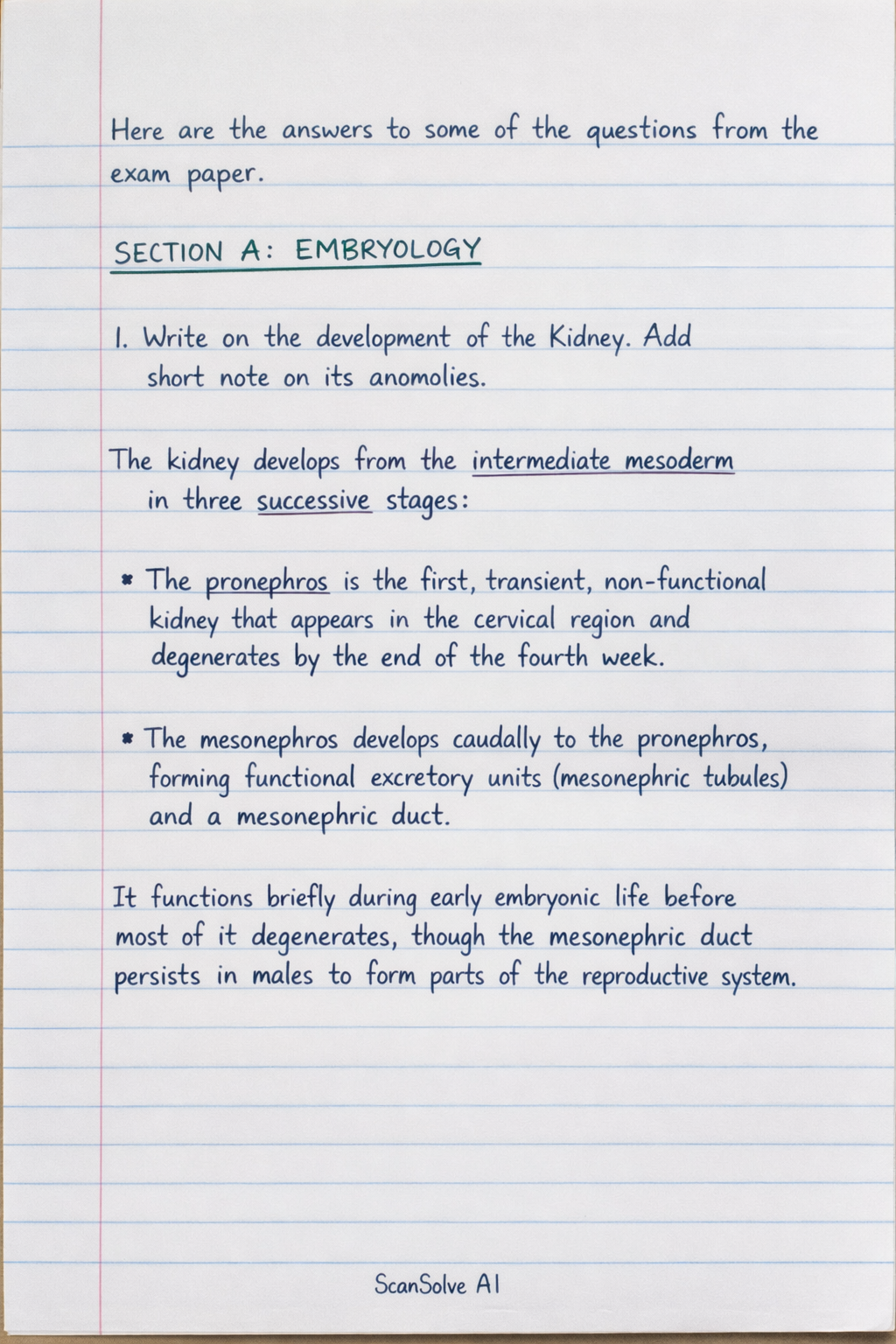
Here are the answers to some of the questions from the exam paper.
SECTION A: EMBRYOLOGY
1. Write on the development of the Kidney. Add short note on its anomalies
The kidney develops from the intermediate mesoderm in three successive stages:
- The pronephros is the first, transient, non-functional kidney that appears in the cervical region and degenerates by the end of the fourth week.
- The mesonephros develops caudally to the pronephros, forming functional excretory units (mesonephric tubules) and a mesonephric duct. It functions briefly during early embryonic life before most of it degenerates, though the mesonephric duct persists in males to form parts of the reproductive system.
- The metanephros is the permanent kidney, appearing in the fifth week. It develops from two sources: the ureteric bud (an outgrowth of the mesonephric duct) and the metanephric mesoderm (or metanephric blastema). The ureteric bud gives rise to the collecting ducts, major and minor calyces, renal pelvis, and ureter. The metanephric mesoderm forms the nephrons (glomerulus, Bowman's capsule, proximal convoluted tubule, loop of Henle, distal convoluted tubule). The interaction between these two tissues is crucial for kidney formation. The kidneys initially form in the pelvic region and ascend to their adult lumbar position.
Anomalies of the Kidney:
- Renal Agenesis: Absence of one or both kidneys. Unilateral agenesis is more common and often asymptomatic, while bilateral agenesis is lethal (Potter sequence).
- Horseshoe Kidney: Fusion of the inferior poles of the kidneys, typically across the midline, forming a U-shape. Its ascent is often arrested by the inferior mesenteric artery.
- Ectopic Kidney: A kidney located in an abnormal position, often in the pelvis, due to failure of normal ascent.
- Polycystic Kidney Disease (PKD): Characterized by numerous cysts in the kidneys. Autosomal dominant PKD (ADPKD) typically manifests in adulthood, while autosomal recessive PKD (ARPKD) is more severe and often presents in infancy or childhood.
SECTION B: HISTOLOGY
1. Describe the histology of the Hyaline cartilage
Hyaline cartilage is the most common type of cartilage, characterized by its glassy, translucent appearance in fresh tissue. It consists of cells called chondrocytes embedded within an extensive extracellular matrix (ECM).
- Chondrocytes: These are mature cartilage cells located in small spaces within the matrix called lacunae. They often appear in clusters of two to eight cells, known as isogenous groups, which arise from the division of a single chondrocyte. Chondrocytes are responsible for maintaining the ECM.
- Extracellular Matrix (ECM): The ECM of hyaline cartilage is composed primarily of Type II collagen fibers and a ground substance rich in proteoglycans (especially aggrecan) and hyaluronic acid. The high water content (up to 80%) bound to the proteoglycans gives hyaline cartilage its resilience and ability to withstand compression. The matrix appears homogeneous because the collagen fibers are very fine and have a refractive index similar to the ground substance, making them difficult to distinguish with a light microscope.
- Perichondrium: Most hyaline cartilage is covered by a layer of dense irregular connective tissue called the perichondrium. The perichondrium has an outer fibrous layer (containing fibroblasts) and an inner chondrogenic layer (containing chondroblasts that can differentiate into chondrocytes, contributing to appositional growth). Articular cartilage, which covers the ends of bones in synovial joints, is an exception and lacks a perichondrium.
Hyaline cartilage provides smooth, low-friction surfaces for joint movement, provides structural support, and allows for flexibility. It is found in the articular surfaces of long bones, costal cartilages, tracheal rings, nasal septum, and parts of the larynx.
SECTION C: GROSS ANATOMY
2. Write on bone markings and classification of joints
Bone Markings:
Bone markings are characteristic surface features on bones that serve various functions, such as points of attachment for muscles, ligaments, and tendons; articulation surfaces for joints; or passages for nerves and blood vessels. They can be broadly categorized into projections (processes) and depressions (openings).
-
Projections/Processes (for muscle/ligament attachment or articulation):
- Head: A prominent, rounded epiphysis (e.g., head of femur).
- Condyle: A rounded articular projection (e.g., medial condyle of femur).
- Epicondyle: A projection superior to a condyle (e.g., medial epicondyle of humerus).
- Trochanter: A very large, blunt, irregularly shaped process (e.g., greater trochanter of femur).
- Tubercle: A small, rounded projection (e.g., greater tubercle of humerus).
- Tuberosity: A large, rounded, roughened projection (e.g., deltoid tuberosity).
- Crest: A narrow ridge of bone (e.g., iliac crest).
- Line: A less prominent ridge than a crest (e.g., gluteal line).
- Spine: A sharp, slender projection (e.g., scapular spine).
- Ramus: An arm-like bar of bone (e.g., ramus of mandible).
-
Depressions/Openings (for passages or articulations):
- Fossa: A shallow, basin-like depression (e.g., olecranon fossa).
- Groove/Sulcus: A furrow (e.g., intertubercular sulcus).
- Foramen: A round or oval opening through a bone (e.g., foramen magnum).
- Meatus/Canal: A canal-like passageway (e.g., external acoustic meatus).
- Sinus: A cavity within a bone, filled with air and lined with mucous membrane (e.g., frontal sinus).
- Fissure: A narrow, slit-like opening (e.g., superior orbital fissure).
Classification of Joints:
Joints (articulations) are classified based on either their structure (the type of material binding the bones together and the presence or absence of a joint cavity) or their function (the amount of movement allowed).
1. Structural Classification:
- Fibrous Joints: Bones are joined by fibrous connective tissue; no joint cavity. Most are immovable.
- Sutures: Immovable joints between skull bones.
- Syndesmoses: Bones connected by a ligament or interosseous membrane, allowing slight movement (e.g., tibiofibular joint).
- Gomphoses: Peg-in-socket fibrous joint (e.g., tooth in alveolar socket).
- Cartilaginous Joints: Bones are united by cartilage; no joint cavity.
- Synchondroses: Bones joined by hyaline cartilage; immovable (e.g., epiphyseal plates, first sternocostal joint).
- Symphyses: Bones joined by fibrocartilage; slightly movable (e.g., pubic symphysis, intervertebral discs).
- Synovial Joints: Bones are separated by a fluid-filled joint cavity. All are freely movable. They typically feature articular cartilage, a joint capsule, synovial fluid, and reinforcing ligaments.
- Types of Synovial Joints: Plane, Hinge, Pivot, Condylar, Saddle, Ball-and-socket.
2. Functional Classification:
- Synarthroses: Immovable joints (e.g., sutures, gomphoses, synchondroses).
- Amphiarthroses: Slightly movable joints (e.g., syndesmoses, symphyses).
- Diarthroses: Freely movable joints (all synovial joints).
Still stuck on this one?
Ask a follow-up, and the answer starts from this question.
More Biology Questions
Handwritten Step-by-Step Solution
Get this as a handwritten image on WhatsApp — like a tutor wrote it for you.
Try on WhatsApp