
We read your photo as:
Slides A and B are histological sections for examination. Using materials provided, a. Stain slide A to demonstrate general tissue morphology b. Stain slide B to demonstrate haemosiderrin c. Comment on the staining reaction
ScanSolve AI Answer
1. Histological sections
-
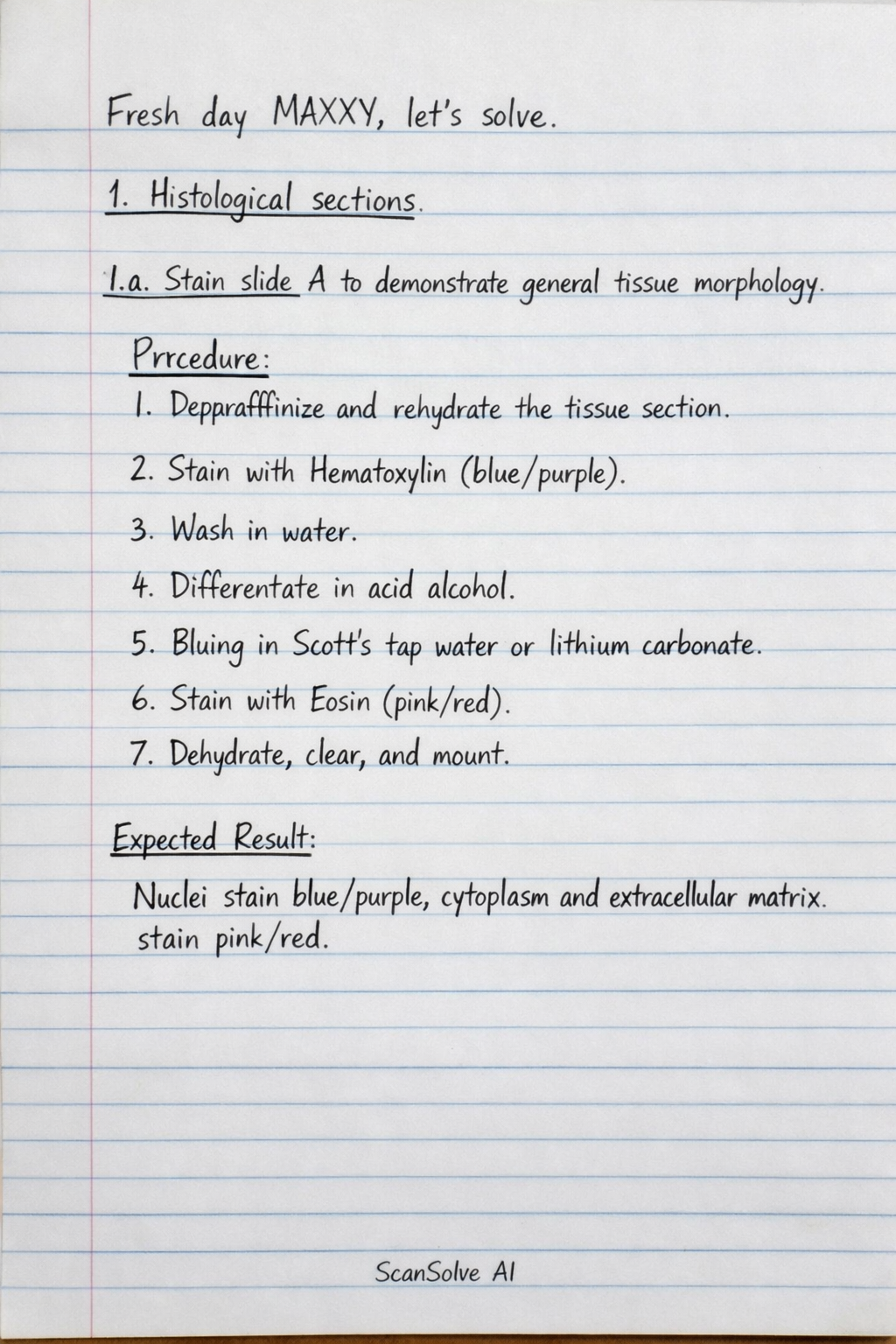
1.a. Stain slide A to demonstrate general tissue morphology
- Procedure: Perform Hematoxylin and Eosin (H&E) staining.
- Deparaffinize and rehydrate the tissue section.
- Stain with Hematoxylin (blue/purple).
- Wash in water.
- Differentiate in acid alcohol.
- Bluing in Scott's tap water or lithium carbonate.
- Stain with Eosin (pink/red).
- Dehydrate, clear, and mount.
- Expected Result: Nuclei stain blue/purple, cytoplasm and extracellular matrix stain pink/red.
- Procedure: Perform Hematoxylin and Eosin (H&E) staining.
-
1.b. Stain slide B to demonstrate haemosiderin
- Procedure: Perform Perls' Prussian Blue stain.
- Deparaffinize and rehydrate the tissue section.
- Incubate in a mixture of potassium ferrocyanide and hydrochloric acid.
- Wash in distilled water.
- Counterstain with Nuclear Fast Red or Safranin.
- Dehydrate, clear, and mount.
- Expected Result: Haemosiderin (ferric iron) deposits stain bright blue. Nuclei stain red (if Nuclear Fast Red is used).
- Procedure: Perform Perls' Prussian Blue stain.
-
1.c. Comment on the staining reaction
- H&E: Hematoxylin is a basic dye that stains acidic components (like nucleic acids in the nucleus) blue/purple (basophilia). Eosin is an acidic dye that stains basic components (like proteins in the cytoplasm and collagen) pink/red (eosinophilia).
- Perls' Prussian Blue: Ferric iron () in haemosiderin reacts with potassium ferrocyanide in an acidic solution to form ferric ferrocyanide, which is an insoluble blue pigment (Prussian blue). This is a histochemical reaction.
2. Urinalysis
-
2.a. Perform urinalysis on urine samples Z & Y and comment on your results
- Procedure:
- Physical Examination: Observe color, clarity, and measure specific gravity.
- Chemical Examination: Dip a reagent strip (dipstick) into the urine, wait the specified time, and compare color changes to the chart for pH, protein, glucose, ketones, blood, bilirubin, urobilinogen, nitrite, and leukocyte esterase.
- Microscopic Examination: Centrifuge a portion of the urine, decant the supernatant, resuspend the sediment, and examine under a microscope for cells (RBCs, WBCs, epithelial cells), casts, crystals, bacteria, yeast, and parasites.
- Comment on Results: Interpret any abnormal findings (e.g., dark yellow urine with high specific gravity and positive glucose suggests dehydration and diabetes; cloudy urine with positive leukocyte esterase and nitrites suggests UTI).
- Procedure:
-
2.b. Carry out Benedict's test on samples Z & Y
- Procedure:
- Add 5 mL of Benedict's reagent to a test tube.
- Add 8 drops (0.5 mL) of urine sample to the reagent.
- Heat the mixture in a boiling water bath for 3-5 minutes.
- Observe the color change.
- Expected Result: A color change from blue to green, yellow, orange, or brick-red precipitate indicates the presence of reducing sugars. The intensity of the color/precipitate indicates the concentration.
- Procedure:
-
2.c. Significance of the presence of (i) reducing substances (ii) protein in urine.
- (i) Reducing substances: The most common reducing substance in urine is glucose, indicating diabetes mellitus. Other reducing sugars like galactose (galactosemia) or fructose (fructosuria) can also be present. Non-sugar reducing substances like ascorbic acid can also give a false positive.
- (ii) Protein in urine (Proteinuria): Indicates potential kidney damage (glomerular or tubular disease), urinary tract infection, hypertension, pre-eclampsia in pregnancy, or other systemic diseases affecting renal function.
-
2.d. What precautions must you take to avoid producing erroneous results.
- Proper Sample Collection: Use clean, dry containers; collect midstream urine; ensure timely delivery to the lab.
- Timely Analysis: Analyze urine within 1-2 hours of collection or refrigerate to prevent bacterial overgrowth and degradation of components.
- Reagent Quality Control: Use fresh, unexpired reagents and reagent strips; store them correctly.
- Proper Technique: Follow manufacturer's instructions for dipstick use (e.g., dipping time, reading time); use appropriate volumes for chemical tests.
- Microscope Calibration: Ensure the microscope is properly calibrated and maintained.
3. Blood samples (Q, R, S)
-
3.a. Determine ABO and Rhesus groups of the samples
- Procedure (ABO Forward Grouping):
- Place drops of anti-A, anti-B, and anti-D sera on a slide or in tubes.
- Add a drop of the patient's red cell suspension to each.
- Mix and observe for agglutination.
- Procedure (ABO Reverse Grouping):
- Place drops of known A cells and B cells on a slide or in tubes.
- Add a drop of the patient's plasma/serum to each.
- Mix and observe for agglutination.
- Procedure (Rh D Grouping):
- Use anti-D reagent with patient's red cells.
- Observe for agglutination. Confirm negative results with a weak D test if necessary.
- Interpretation: Agglutination with anti-A indicates A antigen; with anti-B indicates B antigen. Agglutination with anti-D indicates Rh positive. Reverse grouping confirms ABO type by detecting anti-A and/or anti-B antibodies in plasma.
- Procedure (ABO Forward Grouping):
-
3.b. Determine the PCV of sample Q and comment on your result.
- Procedure:
- Fill a capillary tube with anticoagulated blood (sample Q).
- Seal one end of the capillary tube.
- Centrifuge the tube in a microhematocrit centrifuge for 5 minutes.
- Read the PCV using a microhematocrit reader.
- Comment on Result: Compare the obtained PCV value to the normal reference range (e.g., 0.40-0.54 L/L for adult males, 0.37-0.47 L/L for adult females). A value below the range indicates anemia, while a value above the range indicates polycythemia.
- Procedure:
-
3.c. Make a thin film of sample R and stain for WBC differential count.
- Procedure (Thin Film Preparation):
- Place a small drop of blood (sample R) on one end of a clean glass slide.
- Using a spreader slide held at a 30-45 degree angle, push the drop of blood along the slide to create a thin film with a feathered edge.
- Allow the film to air dry.
- Procedure (Staining - e.g., Leishman stain):
- Fix the dried film with undiluted Leishman stain.
- Add an equal volume of buffered distilled water to the stain, mix, and allow to stand.
- Wash gently with buffered distilled water.
- Allow to air dry.
- Procedure (WBC Differential Count):
- Examine the stained film under oil immersion objective.
- Identify and count 100-200 white blood cells (neutrophils, lymphocytes, monocytes, eosinophils, basophils) in the monolayer area.
- Report the percentage of each type.
- Expected Result: Different WBC types will be visible with characteristic morphology and staining patterns (e.g., neutrophils with multi-lobed nucleus and pink granules, lymphocytes with large nucleus and scant cytoplasm).
- Procedure (Thin Film Preparation):
4. Microbiology
-
4.a. Prepare a MacConkey agar plate
- Procedure:
- Weigh the appropriate amount of dehydrated MacConkey agar powder according to the manufacturer's instructions.
- Suspend the powder in the specified volume of distilled water in a flask.
- Heat the suspension gently with frequent agitation to dissolve the powder completely.
- Autoclave at for 15 minutes.
- Cool the medium to approximately .
- Pour the cooled medium into sterile Petri dishes on a level surface.
- Allow the agar to solidify completely.
- Store inverted at until use.
- Procedure:
-
4.b. i) Perform Gram stain procedure on samples J and L
- Procedure:
- Prepare a thin smear of the sample (J or L) on a clean glass slide and air dry.
- Heat fix the smear by passing it through a flame a few times.
- Flood the smear with Crystal Violet (primary stain) for 1 minute.
- Rinse gently with water.
- Flood with Gram's Iodine (mordant) for 1 minute.
- Rinse gently with water.
- Decolorize with Acetone-Alcohol (decolorizer) for 10-20 seconds until the runoff is clear.
- Rinse immediately with water.
- Flood with Safranin (counterstain) for 1 minute.
- Rinse gently with water, air dry, and examine.
- Procedure:
-
4.b. ii) Examine samples J and L microscopically and report your findings
- Expected Findings:
- Gram-positive bacteria: Appear purple/blue (e.g., Gram-positive cocci in clusters or chains, Gram-positive bacilli).
- Gram-negative bacteria: Appear pink/red (e.g., Gram-negative bacilli, Gram-negative cocci).
- Report morphology (cocci, bacilli, spirilla), arrangement (clusters, chains, pairs), and relative numbers. Also note presence of other elements like WBCs, epithelial cells, or yeast.
- Expected Findings:
-
4.c. Inoculate either sample J or L, on the MacConkey agar plate you prepared and leave on the bench
- Procedure:
- Using a sterile inoculating loop, pick a small amount of the chosen sample (J or L).
- Streak the sample onto the surface of the MacConkey agar plate using the quadrant streaking method to obtain isolated colonies.
- Incubate the plate inverted at for 18-24 hours.
- Purpose: MacConkey agar is selective for Gram-negative bacteria and differential for lactose fermenters (pink/red colonies) versus non-lactose fermenters (colorless colonies).
- Procedure:
5. Parasitology/Routine tests
-
5.a. Examine and carry out routine tests on stool Sample P
- Procedure:
- Macroscopic Examination: Observe color, consistency (formed, soft, watery), presence of blood, mucus, adult worms, or proglottids.
- Microscopic Examination (Wet Mount):
- Prepare a saline wet mount: Mix a small portion of stool with 0.85% saline on a slide, cover with a coverslip.
- Prepare an iodine wet mount: Mix a small portion of stool with Lugol's iodine on a slide, cover with a coverslip.
- Examine both mounts under low and high power objectives for ova, cysts, trophozoites of parasites, WBCs, RBCs, yeast, and undigested food fibers.
- Chemical Examination (Occult Blood): Perform a fecal occult blood test (e.g., guaiac test) to detect hidden blood.
- Expected Findings: Report presence/absence of parasites (e.g., Ascaris ova, Giardia cysts), blood, mucus, and other abnormal findings.
- Procedure:
-
5.b. Examine and carry out routine tests on urine sediment N.
- Procedure:
- Mix the urine sample N thoroughly.
- Pour 10-12 mL of urine into a conical centrifuge tube.
- Centrifuge at 400-500 RCF for 5 minutes.
- Carefully decant the supernatant, leaving approximately 0.5-1 mL of urine with the sediment.
- Resuspend the sediment by gently tapping the bottom of the tube.
- Transfer a drop of the resuspended sediment onto a clean glass slide, cover with a coverslip.
- Examine under low power (10x) and high power (40x) objectives.
- Expected Findings: Report the presence and quantity of:
- Cells: Red blood cells (RBCs), white blood cells (WBCs), epithelial cells (squamous, transitional, renal tubular).
- Casts: Hyaline, granular, cellular (RBC, WBC, epithelial), waxy, fatty casts.
- Crystals: Uric acid, calcium oxalate, triple phosphate, cystine, etc.
- Other: Bacteria, yeast, parasites (e.g., Trichomonas vaginalis), mucus threads.
- Procedure:
-
5.c. Prepare a thick film from blood sample M. Using materials provided, stain your slide for examination for parasites.
- Procedure (Thick Film Preparation):
- Place a large drop of blood (sample M) in the center of a clean glass slide.
- Using the corner of another slide or an applicator stick, spread the blood in a circular motion to form a thick, opaque film about the size of a dime.
- Allow the film to air dry completely in a horizontal position (do not heat fix).
- Procedure (Staining - e.g., Giemsa stain):
- Place the dried thick film in undiluted Giemsa stain for 10-15 minutes (this lyses RBCs and stains parasites).
- Rinse gently with buffered distilled water.
- Allow to air dry vertically.
- Examination for Parasites: Examine the stained thick film under oil immersion objective for the presence of blood parasites, particularly malaria parasites (trophozoites, schizonts, gametocytes). The RBCs will be lysed, leaving only WBCs and any parasites.
- Procedure (Thick Film Preparation):
Still stuck on this one?
Ask a follow-up, and the answer starts from this question.
More Biology Questions
Handwritten Step-by-Step Solution
Get this as a handwritten image on WhatsApp — like a tutor wrote it for you.
Try on WhatsApp